Call us

+91-9240214610 (Bandra)
+91-9240214611 (Kandivali)

  • JAS-ANZ Accredited Certification
  • ICS Certified
  • NABH Certified Eye Hospital in Mumbai
Ojas Eye Hospital in Bandra, Kandivali, Mumbai

Tag: Glaucoma treatment in Mumbai

Which Option Offers the Best Glaucoma Treatment in Mumbai? Drops, SLT Laser or MIGS

Glaucoma increases eye pressure and gradually damages the optic nerve. It is one of the leading causes of irreversible vision loss worldwide. It develops silently, often without symptoms, until severe optic nerve damage has already occurred.

Many people manage it with medicines or simple office-based lasers. When these are not enough, surgery can help lower eye pressure and prevent further vision loss. Surgery won’t restore lost sight but can protect remaining vision. The right treatment depends on your glaucoma type, pressure levels, overall health and whether you’re also planning cataract surgery. Regular follow-ups are essential with every option.

In this article, we will discuss eye drops, SLT laserand MIGS to help you choose the best glaucoma treatment in Mumbai, and for specialized glaucoma care, why to visit Ojas Eye Hospital in Mumbai.

Why Do We Treat Glaucoma?

Glaucoma gradually damages the optic nerve due to elevated intraocular pressure (IOP). Since patients often don’t feel pain or notice early symptoms, treatment focuses on lowering eye pressure to a “target pressure” determined by the glaucoma specialist. This target pressure helps slow or prevent further vision loss.

Because early detection and timely treatment are essential, understanding the different glaucoma treatment options in Mumbai is important to protecting long-term vision. Today, glaucoma care has evolved significantly. Modern treatments help reduce eye pressure using approaches tailored to the patient’s specific type and stage of glaucoma. Whether through eye drops, SLT laser in Mumbai, or MIGS surgery in Mumbai, ophthalmologists now have more effective and safer solutions to manage the disease at every stage.

The right treatment depends on:

  • Type of glaucoma
  • Current eye pressure
  • Extent of optic nerve damage
  • Patient’s age and lifestyle
  • Response to previous medications

Eye Drops: The First Step for Many Patients

For early or mild glaucoma, medicated eye drops are often the first recommended treatment. They work by either reducing fluid production or improving fluid drainage.

Eye drops must be used daily and exactly as prescribed to keep eye pressure under control. Missing doses can raise pressure and increase the risk of optic nerve damage. Some patients may require two or more types of drops to reach their target pressure. Possible side effects include redness, burning, dryness or even changes in eye color.

If drops aren’t effective, cause side effects or patients struggle with compliance, glaucoma specialist often recommend laser therapy – particularly Selective Laser Trabeculoplasty (SLT).

SLT Laser: A Safe, Effective, Low-Maintenance Option

Selective Laser Trabeculoplasty (SLT) is one of the most advanced and widely recommended laser treatments for glaucoma. It improves the eye’s natural drainage system, reducing pressure without surgery.

Why SLT is becoming a first-line treatment

SLT is as effective as daily eye dropsand sometimes even better. It reduces dependence on medication and offers long-lasting results.

Advantages of SLT Laser

  • Performed in clinic, no hospitalization needed
  • Minimal recovery time; patients resume activities the same day
  • Safe and repeatable
  • Often reduces or eliminates the need for eye drops
  • Excellent option for patients who forget daily medications

Many patients now prefer SLT Laser in Mumbai as their initial treatment for open-angle glaucoma or ocular hypertension.

MIGS: Minimally Invasive Glaucoma Surgery

When eye drops and SLT aren’t enough, or when cataract surgery is planned – Minimally Invasive Glaucoma Surgery (MIGS) offers a safer, faster-recovery alternative to traditional surgery.

MIGS procedures use tiny micro-stents or microscopic tools to create new pathways or enhance existing drainage routes.

Categories of MIGS procedure

There are three main categories of MIGS procedure, based on how they lower intraocular pressure:

  • Improving natural drainage system of the eye through the trabecular
  • Creating a new drainage pathway for fluid to exit the eye
  • Reducing fluid production in the eye

Benefits of MIGS surgery

  • Significantly lower risk compared to trabeculectomy
  • Shorter surgery time and faster recovery
  • Can be combined with cataract surgery
  • Reduced dependence on long-term medications
  • Suitable for mild to moderate glaucoma

Because of its safety profile and growing success rates, many specialists recommend MIGS surgery in Mumbai earlier in the treatment process.

Traditional Glaucoma Surgery in Mumbai (For Advanced Cases)

If the glaucoma is severe or pressure needs to be lowered dramatically, traditional surgeries offer stronger pressure reduction:

  • Trabeculectomy
  • Tube shunt surgery

These procedures are highly effective but require longer recovery and carry more risks, so they are reserved for advanced or rapidly worsening glaucoma cases.

Why Regular Follow-Up Is Essential?

Regardless of the treatment chosen, glaucoma must be monitored lifelong. Patients often don’t feel symptoms even as damage progresses, making follow-ups essential for maintaining safe eye pressure.

Which Glaucoma Treatment Works Best – Eye Drops, SLT Laser or MIGS?

Glaucoma treatment depends on eye pressure levels and disease severity. Eye drops help early cases, SLT laser offers a safe, low-maintenance alternative and MIGS suits patients needing stronger pressure control or combining treatment with cataract surgery. Your glaucoma specialist decides the best option based on your condition.

Why Book Your Appointment at Ojas Eye Hospital?

When it comes to glaucoma care, choosing the right hospital and surgeon can protect your vision for life. Ojas Eye Hospital is recognized as the best glaucoma eye hospital in Mumbai, offering advanced diagnostic tools, world-class facilities and highly experienced glaucoma specialists.Whether you need eye drops, SLT laser therapy or advanced MIGS surgery, Ojas ensures you receive the best glaucoma treatment in Mumbai tailored to your condition.

We help keep your vision safe.

10 habits to avoid glaucoma risk: Expert Glaucoma Treatment in Mumbai

Glaucoma, often called the “silent thief of sight,” is a progressive eye disease that damages the optic nerve, leading to irreversible vision loss if left untreated. Glaucoma usually develops without noticeable symptoms until significant damage has occurred. Nowadays, advancements in medical science have made treatment options available, but prevention is still better than cure.

This blog highlights 10 daily lifestyle habits to avoid in order to reduce your risk of developing glaucoma and preserve your vision for the long term.

Below are 10 habits you should avoid to reduce your risk of glaucoma:

Maintain an Active Lifestyle

A sedentary routine affects your overall health, including your eyes. Regular exercise helps regulate blood flow and intraocular pressure.

Expert tips:

  • Include moderate exercises like walking, yoga, or swimming in your routine.
  • Avoid heavy weightlifting, which can spike eye pressure.

Watch Your Caffeine Intake

Caffeine in large amounts can cause temporary spikes in eye pressure, especially in individuals at risk for glaucoma.

Expert tips:

  • Limit your coffee or energy drink consumption to 1–2 cups daily.
  • Opt for decaffeinated beverages when possible.

Quit Smoking and Cut Down on Alcohol

Smoking introduces harmful chemicals that can damage the optic nerve and elevate your risk of glaucoma. Excessive alcohol can also dehydrate the body, disturbing the eye’s fluid balance and increasing IOP.

Expert tips:

  • Avoid tobacco products completely.
  • Limit alcohol intake to moderate levels.

Avoid Excessive Eye Rubbing

Rubbing your eyes may provide temporary relief but can increase eye pressure and damage delicate eye structures.

Expert tips:

  • Use lubricating eye drops or splash clean water if your eyes feel dry or irritated.
  • If symptoms persist, consult an eye specialist in Mumbai instead of self-medicating.

Eat a Balanced, Eye-Healthy Diet

Unhealthy diets rich in processed food, sugar, and excess salt contribute to systemic conditions like diabetes and hypertension, and both are linked to glaucoma.

Expert tips:

  • Eat a balanced diet rich in leafy greens, omega-3 fatty acids, carrots, citrus fruits, and antioxidant-rich foods.
  • Stay hydrated to maintain proper eye fluid balance.

Don’t Skip Regular Eye Check-ups

Glaucoma develops slowly and is often undetected until significant vision loss occurs. That’s why routine eye check-ups are essential for early detection.

Expert tips:

  • Adults over 40 should get complete eye exams every 1–2 years.
  • Those with a family history of glaucoma should start earlier.

Take Eye Injuries Seriously

Even minor eye trauma can lead to secondary glaucoma. Protective measures are essential when engaging in sports or hazardous tasks.

Expert tips:

  • Use protective eyewear during contact sports, when working with machinery, or during risky tasks.
  • If you experience an eye injury, consult an eye specialist in Mumbai immediately.

Poor Sleep and High Stress

Lack of sleep and constant stress may contribute to systemic hypertension, which in turn elevates your IOP.

Expert tips:

  • Practice relaxation techniques such as meditation or breathing exercises.
  • Ensure 7–8 hours of quality sleep each night.

Say No to Self-Medication

Many people misuse over-the-counter eye drops without proper diagnosis. Some of these may contain steroids that can worsen or trigger glaucoma.

Expert tips:

  • Never use eye medications without a prescription.
  • Consult a certified ophthalmologist in Mumbai for any eye discomfort or visual changes.

Limit Excessive Screen Time

Spending long hours on smartphones, laptops, or TVs can lead to digital eye strain and elevated intraocular pressure (IOP). This is especially concerning for individuals already at risk of glaucoma.

Expert tips:

  • Follow the 20-20-20 rule: Every 20 minutes, look 20 feet away for 20 seconds.
  • Use anti-glare screens and take frequent breaks.

Why Early Prevention Matters

Glaucoma can lead to permanent blindness if not detected and managed early. While there’s no guaranteed way to avoid it entirely, taking preventive steps through lifestyle adjustments and regular check-ups drastically lowers your risk.

Book Your Consultation with the Best Glaucoma Specialist in Mumbai

When it comes to your vision, we can only trust the most experienced hands. Ojas Eye Hospital in Mumbai is widely recognized for offering the best glaucoma surgery in Mumbai, led by the best glaucoma specialist in Mumbai.

Book your appointment at the best eye hospital in Mumbai for expert care and long-term relief.

How Can Stress Management Improve Glaucoma Treatment in Mumbai?

Recent researches have revealed a significant link between psychological stress and raised intraocular pressure (IOP) in individuals with primary open-angle glaucoma (POAG). This research highlights the importance of stress management for glaucoma patients and provides valuable details about how stress could potentially lead to irreversible vision loss if not managed properly.

What is Glaucoma?

Glaucoma is a progressive eye disease characterized by optic nerve damage that can result in vision impairment or even blindness. Elevated intraocular pressure (IOP) is a major risk factor for worsening glaucoma, significantly affecting a patient’s quality of life. The most common form of glaucoma is the open-angle glaucoma (POAG), which is often managed by controlling IOP, that is the fluid pressure inside the eye.

How Is Stress and Intraocular Pressure (IOP) Linked?

Traditionally, glaucoma treatments focus on lowering intraocular pressure (IOP) through medications, laser therapy, or surgery. However, stress has been observed to temporarily raise IOP, even in individuals without glaucoma. This has made doctors to investigate whether stress has a deeper impact on glaucoma patients.

Could Stress Worsen Glaucoma?

Yes, repeated stress exposure is believed to impair IOP control in glaucoma patients, potentially increasing the risk of disease progression. Therefore, managing both physical and emotional health is essential as part of a complete glaucoma treatment plan.

How to Manage Stress for Better IOP Control?

Incorporating stress relaxation methods into daily routines may help glaucoma patients manage IOP more effectively. Consider the following methods:

  • Mindfulness meditation: Helps calm the mind and reduce stress hormones.
  • Deep breathing exercises: Promotes relaxation and lowers blood pressure.
  • Physical activities or tai chi: Combines physical movement with mental well-being.
  • Cognitive-behavioral therapy (CBT): Provides coping strategies for managing stress and anxiety.

Why Timely Treatment of Glaucoma Matters?

Glaucoma is often called the “silent thief of sight” because it progresses without noticeable symptoms until significant vision loss occurs. Regular screenings and timely intervention are essential for preserving vision and quality of life.

Act Today, Visit Ojas for the Top-Quality Glaucoma Treatment in Mumbai

Ojas Eye Hospital is known for providing the best glaucoma treatment in Mumbai, combining the most advanced procedures with expert care. The hospital has some of the best eye specialist in Mumbai and the best eye surgeon in Mumbai, with extensive experience in diagnosing and treating complex eye conditions.

Having state-of-the-art equipment for vision correction and glaucoma surgeries makes Ojas -Eye Hospital a leading destination for top-quality glaucoma treatment in Mumbai.

Your vision is valuable, and protecting it requires expert care and timely attention. Whether you’re seeking the best glaucoma treatment in Mumbai or advanced surgical solutions, Ojas – Eye Hospital is your trusted choice. Book your appointment today at the best eye hospital in Mumbai and take a step toward a brighter, healthier future for your eyes.

Everything you need to know about pigmentary glaucoma

– Dr Niteen Dedhia, Dr Rushita Kamdar

Pigmentary glaucoma is caused by pigment dispersion syndrome

Pigmentary glaucoma is a type of glaucoma brought on by pigment dispersion syndrome (PDS). PDS occurs when pigment granules (the pigments that give your eyes their colour) flake off the iris and end up in the clear fluid of your eye, known as the aqueous humour (release of execessive pigment in the anterior segment of the eye.)

Another possibility is that your iris and lens are in contact, so that when you look around and focus your eyes on different text or objects, your iris rubs against the lens. As a result, some of the iris’s pigment flakes off before it enters the aqueous humour. Mild pigment dispersion, on the other hand, is a normal part of the ageing process and is nothing to be concerned about as long as your eye pressure is normal.

According to experts, approximately one-third of PDS patients develop pigmentary glaucoma. Glaucoma develops when the pigment in the aqueous humour clogs the drainage system of the eye, causing pressure to build up inside the eye. Some people with PDS can tolerate extra pigment in their aqueous humour and it drains normally. On the other hand, some people find that it significantly raises their eye pressure, putting them at risk of pigmentary glaucoma. PDS affects both men and women equally, but men are three times more likely than women to develop pigmentary glaucoma. Furthermore, 80% of people with PDS are myopic, indicating that being short-sighted is a significant risk factor for the condition.

When your eye pressure is higher than it should be for an extended period of time, you run the risk of damaging your optic nerve. Black spots appear across your visual field as a result of optic nerve damage, as does loss of colour vision. This happens when the optic nerve fibres die as a result of high eye pressure. Often, people notice that glaucoma affects their peripheral vision first. However, by the time you notice it, it is too late to recover any lost vision.

What are the signs and symptoms of pigment dispersion syndrome?

PDS is often asymptomatic and can only be detected during a routine eye exam. It causes pigmentary glaucoma, which is most commonly diagnosed in people in their twenties, thirties, and forties. This is significantly younger than the average age at which other types of glaucoma develop and are diagnosed. Although PDS is often asymptomatic, the following symptoms may occur:

  • Hazy vision
  • When looking at bright lights, you may notice halo effects.
  • Eye ache
  • Swelling of the cornea

If you notice any of these symptoms, or if you have pigmentary glaucoma symptoms such as black spots in your visual field, see your optician as soon as possible. There are treatment options available, and even if your vision has not been affected yet, regular monitoring is essential to determine if and when treatment is required.

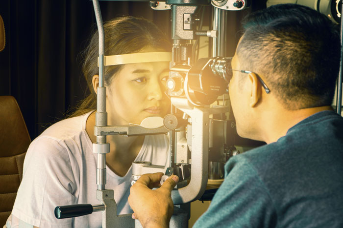
Ocular examination – careful slit lamp examination is important to look for the presence of pigment granules. Iris should be examined for any defect. Lens should be examined for the presence of pigment on the anterior segment .angle of anterior chamber should be examined for any pigment deposition. intraocular pressure should be measured.

How can pigment dispersion syndrome and pigmentary glaucoma be treated?

If pigment dispersion syndrome is detected during a routine eye exam and you have no symptoms, you probably won’t need any medication or treatment right away. Mild pigment dispersion, as mentioned briefly in the first paragraph, is a normal part of the ageing process of the eye. Only when your eye pressure rises and you begin to show signs of glaucoma should you take action.

Because vigorous exercise can cause pigment release, some patients with a high number of pigment deposits in their eyes are advised to avoid vigorous exercise whenever possible, or to take medication to lower their eye pressure before engaging in a strenuous workout. One of the main treatment options for people with pigment dispersion syndrome is medication to lower their eye pressure, which usually comes in the form of medicated eye drops.

Laser treatment may be recommended in severe cases of PDS or when your eye pressure becomes so high that it affects your optic nerve. In this type of surgery, the laser is used to make a small hole in your iris to treat pigmentary glaucoma. This does not improve your vision; instead, it helps to shift the anatomy of your eye so that there is less rubbing between the iris and lens, resulting in less pigment release.

If you suspect you have PDS or are concerned about symptoms of glaucoma, see us for a check-up at Ojas Eye Hospital. We will be able to advise you on the best course of action and arrange for any sight-saving treatment that is required.

Contact Lens Sensor In Monitoring 24-Hour IOP

Common Eye Myths And Myth Busters

Glaucoma is a condition in which increased intraocular pressure leads to alteration in the visual function. The symptoms of glaucoma are believed to be caused due to pressure on the posterior part of the eye, especially the lamina cribrosa, which is a part of the back of the eye where the sclera gives way to the retinal ganglion cell axon to leave the eye.

It is essential for the people undergoing treatment for glaucoma to get their IOP (INTRA OCULAR PRESSURE) checked at frequent intervals to evaluate the efficacy of the treatment. Almost all the patients undergo a daytime intraocular pressure measurement at the doctors office due to its convenience. However, the efficacy of the daytime measurement of IOP is under serious debate as people who have well-managed daytime intraocular pressure also complain about the development of the symptoms of progression of glaucoma including visual field defects. Intraocular pressure does not remain the same throughout the day and measuring the extent of fluctuation during 24 hours is the mainstay in deciding the treatment strategy for controlling this risk factor.

Various studies have indicated that patients with low IOP have their disease progressing leading to reduced eyesight. This is supposed to be due to variation in IOP which makes optic nerve vulnerable to increased mechanical stress and strain. The stress on the optic nerve is measured far better through 24- hour variability rather than measuring the IOP only during office hours.

Common methods used for measuring IOP include handheld applanation tonometry, Goldman applanation tonometry (GAT), and pneumotonometry. These methods are largely used in day time settings and are not implemented in 24-hour IOP measurement because they are time-consuming and inconvenient to the patient. Various ambulatory methods for measuring 24-hour IOP have been developed. These methods include permanent IOP monitoring, self-tonometry, and temporary IOP monitoring.

The researchers found that IOP is a principal and only modifiable risk factor for the progression of glaucoma. However, the lack of effective methods for IOP data collection reduces the efficacy of strategies for managing IOP. Various decisions related to the management and surgery for glaucoma are based on ambiguous and unauthenticated IOP data leading to poor outcomes. The IOP characteristics such as mean, peak and fluctuations in IOP should be accurately measured to improve therapeutic efficacy.

A study was done to determine the use of a contact lens sensor device in measuring the 24-hour IOP in the patients who had undergone various interventions in the management of glaucoma. The contact lens sensor device works on the mechanism that fluctuations in intraocular pressure change the curvature of cornea which is measured by this device. This device provides a 24-hour data related to changes in IOP. The study also determines which intervention is the most effective in providing minimum IOP fluctuations.

The study includes 30 centers from sixteen countries and a total of 182 patients were involved. The number of eyes undergone interventions was 182. IOP in 60 eyes was managed through topical medications, incisional surgery was done in 53 eyes, and 69 eyes were with laser trabeculoplasty. The ocular dimensional profile was measured before and after the interventions through CLS (CONTACT LENS SENSOR )

Patients with ocular hypertension, open-angle glaucoma and who are glaucoma suspects were included in the study with a provison that they must have reliable and sufficient CLS readings. Patients who do not have complete and sufficient readings were excluded from the study. Various statistical tools were used during the study such as linear regression and Benjamini-Hochberg approach. Spearman rank correlation coefficient was used to analyze the relationship between changes in CLS parameters and GAT ( GOLDMAN APPLANATION TONOMETER) IOP.

The study concludes that all three interventions, viz, the medications, laser trabeculoplasty and, incisional surgery were effective in managing the IOP. In the patients with incisional surgery, the IOP before and after the intervention was 21.4 mm Hg and 14.7 mm Hg respectively, indicating a mean reduction of 6.8 mm Hg. In the laser trabeculoplasty patients, there was a mean reduction of 2.7 mm Hg with before and after the intervention, IOP measured 17.2 mm Hg and 14.6 mm Hg respectively. The topical medications lower the IOP by 0.97 mm Hg with measured IOP before intervention 18.1 mm Hg and after intervention 17.1 mm Hg.

Incisional surgery was found to be significantly effective on 20 CLS parameters as compared to the other two methods while on one parameter, laser trabeculoplasty was more effective than topical medications. Incisional surgery was found to be predictive for most of the changes in CLS parameters. The researchers indicated that contact lens sensor is an effective method to monitor the 24-hour IOP fluctuations as compared to GAT which is mostly used for day time IOP measurement. The researchers also found that sensitivity was more in nocturnal CLS parameters as compared to diurnal parameters. The authors concluded that this device is effective in measuring the efficacy of the treatment.

A multicenter, retrospective cohort study was conducted on 445 patients at 50 centers in 13 countries. The study also advocates the used of CLS in measuring the IOP. The study indicates that the 24-hour CLS measurements may be associated with previous rates of progression of glaucoma. It is a more convenient and effective method than Goldman which requires multiple time measurements during office hours. The CLS, according to the study, is a useful tool in analyzing the future progression of glaucoma.

In a prospective, cross-sectional study done on forty treated patients, it was found that CLS parameters were related to the field of progression in treated eyes. The device is effective, especially in high-risk eyes, in monitoring IOP while receiving treatment.

Reference:

Chun Hing Ho and Jasper K. W. Wong, Role of 24-Hour Intraocular Pressure Monitoring in Glaucoma Management, Journal of Ophthalmology, Volume 2019

Anastasios G. Konstas, Malik Y. Kahook et al, Diurnal, and 24-h Intraocular Pressures in Glaucoma: Monitoring Strategies and Impact on Prognosis and Treatment, November 2018, Volume 35, Issue 11, pp 1775–1804

Cutolo CA, De Moraes CG, The Effect of Therapeutic IOP-lowering Interventions on the 24-hour Ocular Dimensional Profile Recorded with a Sensing Contact Lens, J Glaucoma. 2019 Mar;28(3):252-257

De Moraes CG, Mansouri K et al, Association Between 24-Hour Intraocular Pressure Monitored with Contact Lens Sensor and Visual Field Progression in Older Adults with Glaucoma, JAMA Ophthalmol. 2018 Jul 1;136(7):779-785

De Moraes CG, Jasien JV, Visual Field Change and 24-Hour IOP-Related Profile with a Contact Lens Sensor in Treated Glaucoma Patients. Ophthalmology. 2016 Apr;123(4):744-53

Dunbar GE, et al. The Sensimed Triggerfish contact lens sensor: efficacy, safety, and patient perspectives. Clin Ophthalmol. 2017; 11: 875–882.

Glaucoma

Digital Eye Strain

A new innovation in Glaucoma treatment. The Beacon Aqueous Microshunt (MicroOptix) –  the brain child of David Brown, MD, 20 years in development-is 3.1 mm in length with an external flange width of 7 mm. It is implanted into a paracentesis-like incision, allowing aqueous to flow through a central channel.

 Mitigation against retrograde flow of pathogens would be a concern for such an idea. However, Dr. Riedel said that this is prevented 1 ) by shear stress and 2) by the polyethylene glycol material that composes the central layer of the three-layer device. Polyethylene glycol is considered the gold standard of anti-bio fouling materials. It is super hydrophilic, and it prevents adherence of protein and bacteria to the channel wall, Dr. Riedel explained.

In a porcine animal model followed to 6 months, Dr. Riedel noted no infections and significant, persistent IOP lowering. An aggressive bacterial challenge with the device on eight animals did not result in eye infection, Dr. Riedel said.

Preliminary data from an early bird study in the U.S. and E.U. has shown a mean IOP ( eye pressure ) of 13 mm Hg at 6 months, compared to a baseline mean of 27 mm Hg that was medicated with no washout of patients. Eighty-seven percent of patients showed a 20% reduction in pressure compared to baseline and all patients except one were off all medications postop. Most patients, Dr. Riedel said, were vet or extremely comfortable at 6 months.

In terms of safety, there were no infections or uveitis, no dislocations or device migration, and no sight-threatening complications. There were there device blockages, two of which resolved completely with no or minimal in tervention. The third was remove for persistent blockage.

Dr. Riedel said investigators will soon conduct a prospective, multicenter study to evaluate safety and efficacy in refractory glaucoma.

Ojas Eye Hospital A Center of Excellence for Contoura Vision, Femto Bladefree Lasik in Mumbai, India.