Call us

+91-9240214610 (Bandra)
+91-9240214611 (Kandivali)

  • JAS-ANZ Accredited Certification
  • ICS Certified
  • NABH Certified Eye Hospital in Mumbai
Ojas Eye Hospital in Bandra, Kandivali, Mumbai

Tag: Retinal Detachment

Can Atropine Eye Drops Help Control Myopia at Every Age?

Myopia, also called short-sightedness, is a very common eye problem today. People with myopia can see nearby objects clearly, but distant things like road signs, classroom boards or TV screens appear blurry. This condition usually starts in childhood and often increases as the child grows.

In recent years, myopia has become more common because children spend more time on mobile phones, tablets and computersand less time outdoors. This makes early eye check-ups and timely treatment extremely important.

At Ojas Eye Hospital, myopia is managed with modern, proven treatments that focus not just on clear vision today but also on protecting eye health in the future.

Why Myopia Should Not Be Ignored?

Many parents think myopia only means wearing stronger glasses every year. But if myopia keeps increasing without control, it can lead to serious eye problems later in life.

Uncontrolled myopia may increase the risk of:

  • Retinal detachment
  • Glaucoma
  • Early cataracts
  • Myopic macular degeneration

These conditions can affect vision permanently. That is why eye specialists strongly recommend early treatment to slow down myopia progression, especially in children.

What Are Atropine Eye Drops?

Atropine eye drops are one of the most trusted and widely researched treatments used to slow down the progression of myopia in children. These drops are given in very low doses and are usually applied once a day, often at night.

Atropine works by relaxing the eye’s focusing muscles and slowing the growth of the eyeball. Since myopia worsens mainly due to excessive eye elongation, controlling this growth helps keep the power stable for a longer time. Several studies have shown that low-dose atropine is safe and effective when used under proper medical supervision.

Do Atropine Drops Work for All Age Groups?

Atropine drops are most effective in children, especially those between early school age and teenage years. During this time, the eyes are still growing and myopia tends to progress faster. Starting treatment early can make a big difference in long-term vision.

Children who benefit the most from atropine treatment usually:

  • Show rapid increase in spectacle power
  • Have a family history of high myopia
  • Spend long hours on screens
  • Spend limited time outdoors

For teenagers, atropine can still help, but the response may vary depending on eye growth.

In adults, the eyes have mostly stopped growing. Because of this, atropine drops usually do not show strong results in slowing myopia. Adults may benefit more from other vision correction recommended by an eye specialist.

Other Myopia Management Options

Myopia control is not the same for everyone. At Ojas Eye Hospital, doctors carefully examine the eyes and suggest the most suitable treatment based on age, lifestyle and eye condition.For those looking for advanced myopia treatment in Mumbai, a personalized plan ensures safetyand long-term results.

Depending on the patient, options may include:

  • Atropine eye drops
  • Special myopia control spectacle lenses
  • Customized contact lenses
  • Orthokeratology (night lenses)
  • Regular vision monitoring

A Personalized Approach to Eye Care

Every eye is different. That is why there is no one treatment for all. At Ojas Eye Hospital, detailed eye examinations are done using advanced diagnostic equipment to understand how fast myopia is progressing and what treatment will work best.

Doctors consider:

  • Age of the patient
  • Current eye power
  • Eye health and structure
  • Daily screen usage
  • Family eye history

Based on this, a customized myopia control plan is created, especially for children who need careful long-term monitoring.

Why Book an Appointment at Ojas Eye Hospital in Mumbai?

Atropine eye drops play an important role in controlling myopia when started at the right age and used correctly. While they may not be suitable for everyone, early diagnosis and expert guidance can protect vision for years to come.

Ojas Eye Hospital is known for providing ethical, patient-focused eye care with modern procedures and experienced specialists. Patients receive clear guidance and honest advice for long-term vision health.

Whether you are consulting the best eye specialist in Mumbai or searching for the best eye hospital in Mumbai, Ojas offers care you can rely on.

Healthy eyes begin with the right care at the right time.

Myopia Control for Kids – What’s Right for My Child: Ortho-K Lenses or Atropine Eye Drops?

Childhood myopia, also known as nearsightedness, is a growing concern for families worldwide. It is a common condition usually diagnosed in young children. More screen time, less outdoor play and increased near work have made blurry distance vision extremely common among children. Glasses help kids see clearly, but they don’t stop myopia from getting worse. That’s why treatments like Ortho-K lenses and atropine eye drops for kids are used to slow its progression.

This article explains how Ortho-K lenses and atropine eye drops for kids work, which children benefit the most and what parents should know before choosing the right option.

What Is Myopia, and Why Does It Progress in Children?

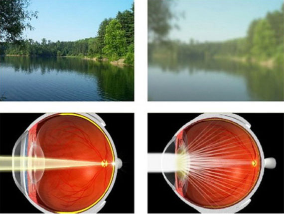
Myopia occurs when the eye grows too long or the front surface (cornea) becomes too curved. This causes light to focus in front of the retina instead of on it, making distant objects appear blurry. In children, myopia usually increases every year because the eyeball continues to grow.

Why Is Myopia Control for Children Important?

If myopia isn’t managed early, it keeps getting worse, leading to frequent prescription changes and a higher risk of serious eye problems. Uncontrolled progression can lead to high myopia, which increases the lifetime risk of:

Managing myopia in children early helps protect their long-term eye health.

Myopia Control Options for Kids

Parents today have two scientifically proven treatments to slow myopia progression:

  • Orthokeratology (Ortho-K) lenses
  • Low-dose atropine eye drops (0.01%–0.05%)

Both options work, but in different ways. Understanding them helps you choose what fits your child’s needs and lifestyle.

Ortho-K Lenses (Orthokeratology)

Ortho-K lenses are special rigid lenses worn only at night. While your child sleeps, the lenses gently reshape the cornea to reduce myopia, meaning your child wakes up with clear vision all day without wearing glasses. When used regularly, Ortho-K lenses can slow myopia progression by nearly 50%, helping lower the risk of serious eye problems in the future.

Benefits of Ortho-K lenses

  • No need for daytime glasses or lenses
  • Excellent for active children, swimmers, athletes
  • Offers freedom from daytime eyewear
  • Strong myopia-control effect
  • Works especially well for children with fast progression

Low-Dose Atropine Eye Drops for Kids

Low-dose atropine (0.01–0.05%) is one of the world’s most widely used treatments for childhood myopia control. These drops relax certain focusing mechanisms and act on receptors in the sclera and retina, helping slow down the eye’s axial growth.

Benefits of Atropine Drops for Kids

  • Easy to use, 1 drop in each eye at bedtime
  • Proven myopia control for children
  • Very low risk of side effects
  • Works for children as young as 4-5 years
  • Ideal for kids who cannot handle lenses

Ortho-K Lenses vs Atropine Eye Drops: Which Is Better for Your Child?

Both treatments are effective, but the best choice depends on factors like age, myopia level, progression speed, eye health and lifestyle.

When Ortho-K May Be the Better Choice

  • Child wants freedom from daytime glasses
  • Active in sports or swimming
  • Moderate myopia
  • Rapid progression
  • Motivated for proper lens care

When Atropine Drops May Be Better

  • Very young children
  • Child cannot handle lenses
  • Parents prefer a simple night routine
  • Early myopia
  • Co-existing dryness or allergies

Best Myopia Control Results

The effect is often strongest when Ortho-K and atropine are used together, especially in children with fast-progressing myopia.

Other Supportive Measures for Myopia Control in children

Along with Ortho-K or atropine, lifestyle changes help reduce progression:

  • 2 hours of outdoor play daily
  • Limit screen time, especially close viewing
  • Maintain proper reading distance
  • Ensure good lighting while reading
  • Take eye breaks every 20 minutes

Why Parents Trust Ojas Eye Hospital for Pediatric Myopia Control in Mumbai

Myopia in children is progressing faster than ever, but modern treatments like Ortho-K lenses and atropine eye drops offer real solutions to slow its progression. With expert guidance from the best ophthalmologist in Mumbai at Ojas Eye Hospital, parents can make informed decisions and give their children the best chance at stable, healthy vision.

Ojas Eye Hospital provides advanced myopia control for kids, supported by experienced pediatric ophthalmologists and state-of-the-art technology.

Act today to protect your child’s tomorrow; smart myopia control now can shape a lifetime of strong, stable vision.

Post-Cataract Wavy Vision: Is it Normal or a Sign of Concern?

Cataract extraction is the most commonly performed eye procedure worldwide, followed by an excellent success rate. For most individuals, the aftereffects can be life-changing: clearer vision, vibrant colours, and improved quality of life. However, in some cases, after surgery, the patient may go through some rather unusual events during the recovery time. One of the features that makes many patients somewhat apprehensive after surgery is wavy lines or some kind of distortion that appears post-procedure.

Is this something dangerous, or a natural part of healing? Let us look at what causes wavy vision after cataract surgery and what will be normal or not require Attention. What To Do (those things that can help the best possible improvement) to ensure the best possible recovery after cataract surgery.

What Makes Straight Lines Wavy After Surgery?

High levels of distortion or waviness after cataract surgery bring anxiety to most people, but do not usually mean something serious. The following are the most common reasons identified for the condition:-

Adjusting the Visual System

Replacement of the cloudy cataract lens by the artificial intraocular lens (IOL) requires time for adaptation by the eyes and the brain. During this phase of adaptation, light that is processed differently by the brain can give rise to temporary distortions such as wavy or curved lines. It compensates itself usually within a few days to weeks.

Cystoid Macular Oedema

Sometimes, a slight swelling of the central area of the retina, the part responsible for sharp vision, sometimes occurs. It causes those lines to be wavy or unclear. Cystoid macular oedema is the name of the illness. Treating it with anti-inflammatory eye drops or injections at home resolves it and returns normal vision.

Epiretinal Membrane

Sometimes, a skinny layer of scar tissue develops on the retina’s surface, which causes wrinkling of the macula. It is called an epiretinal membrane, or more commonly, macular pucker. It can create an optical distortion when viewing bent straight lines. Minor degrees of this disorder may not require intervention. More severe cases might require specialised surgery to remove the membrane.

Lens Visual Phenomena

Some patients have some optical side effects caused by the artificial lens implanted during surgery. Halos, glare, or shadowing phenomena are collectively termed dysphotopsia. Most of these symptoms subside as the eye adapts, but a few patients still complain of persistent distortions and need corrective approaches.

Is Wavy Vision Normal or an Alarm Code?

Wavy vision is often not serious in most cases after cataract surgery. It is often just part of the ordinary healing and adaptation process.

Generally Normal

  • Mild distortion for the first few days after surgery.
  • Symptoms are gradually improving over time.
  • No other warning signs such as pain or severe loss of vision, or flashes of light.

When Something is Wrong

  • Getting worse, rather than improving, distortion.
  • Blurred central vision or trouble with reading.
  • Symptoms appear, and new ones develop, like flashes of light, floating spots, or a dark curtain in your vision.
  • Increased redness, pain, or swelling of the eye.

If any of these warning signs occur, there should be an immediate consultation because these may indicate serious complications such as macular swelling, retinal detachment, or even worse things.

What Can Be Done about Persistent Wavy Vision?

The kind of treatment prescribed depends on what causes the disease. Here are some of the common approaches:-

  • Watch & Wait – For mild symptoms that show up a little after surgery, your physician may suggest watchful waiting for several weeks because, in many cases, natural adaptation will cure this issue.
  • Eye Drops or Injections – When macular swelling is found, those anti-inflammatory eye drops or injections into the eye will lessen the fluid and clear the way to recover the good vision.
  • Surgical Intervention – Where an epiretinal membrane is involved, the procedure to be done is vitrectomy, which consists in peeling off the scar tissue. This type of surgery would almost certainly improve distortion, although the time needed for recovery can vary.
  • Corrective Lens Problems – In cases where the artificial lens causes an enduring optical disturbance, lens repositioning, changing to a different lens type, or employing several corrective techniques are among the possible solutions.

Cataract Surgery Recovery—Tips for a Smooth Recovery

  • Follow Post-Operative Instructions: Use the prescribed eye drops according to instructions and attend all appointments.
  • Protect Your Eyes: Do not rub your eyes; wear protective glasses when necessary.
  • Monitor Your Vision: Keep a record of any changes. Instruments like an Amsler grid can be helpful at home to detect early distortions in central vision.
  • Talk to Your Doctor: Report any unusual or more bothersome experiences.

Can Wavy Vision Be Prevented?

Though the cases can never be entirely prevented, pursuing a carefully planned surgery and duly carried out post-operative measures significantly reduces any risk. Patients already having retinal conditions should address this prior to surgery with the ophthalmologist. In some cases, extra measures or adjunctive treatment could be provided to lessen the risk of complications.

When to Seek Urgent Care

Seek immediate care if:-

  • Sudden increase in distortion or blurring.
  • Flashes of light, or a sudden shower of floaters.
  • A curtain or shadow moving across your field of vision.
  • Severe pain in the eye or sudden loss of vision.

These could be the signs of retinal detachment and other emergencies that must be treated promptly to prevent permanent vision loss.

Conclusion

The presence of wavy or distorted vision after cataract surgery can be disconcerting; however, in many cases, it is a transitory phase of adjustment of the eyes to the new lens. Nevertheless, the persistence or aggravation of symptoms must never be brushed away. Early detection and timely treatment of complications can differentiate between a smooth recovery with minor hiccups and prolonged visual impairment.

Why a Retina Test before LASIK Surgery is Essential to Ensure Safety and Success

Introduction

Having LASIK (Laser-Assisted In Situ Keratomileusis) eye surgery – a highly sophisticated, rapid, and efficient vision corrective surgery that provides long-term results for most patients – is the dream of most people afflicted with refractive disorders such as nearsightedness, farsightedness, or astigmatism for clear, glasses-free vision for which proper checkup of their retina, before LASIK surgery is must that guarantees safety, success, as well as overall eye health in the long term.

A retina check-up assists the ophthalmologist in detecting any pre-existing retinal problems, retinal detachment, or damaged retina, which may subsequently cause severe complications following LASIK surgery if not detected. Therefore, a thorough retina check-up prior to LASIK is necessary to avoid vision-threatening complications and achieve a successful outcome of LASIK.

In this article, we will discuss why a retina test is required prior to LASIK eye surgery, what occurs during a retina test, the risks of avoiding the test, and how it provides vision safety in the long run.

What is a Retina Test?

Retina test is an entire eye examination that checks the condition and well-being of the retina (light-sensitive tissue in the back of the eye). The retina is employed to transmit visual data to the brain to enable you to see properly. Any retinal abnormality will result in foggy vision, loss of vision, retinal detachment, or other problems.

Retina testing is conducted by an eye physician (ophthalmologist) with the help of sophisticated diagnostic equipment such as:-

  • Retinal Imaging (OCT – Optical Coherence Tomography).
  • Fundus Photography.
  • Retina Scanning.
  • Dilated Eye Exam.

The test assists in diagnosing early retinal detachment signs, retinal holes, lattice degeneration, or any other retinal condition that could be a risk factor during or after LASIK surgery.

Why is a Retina Test Needed Before LASIK Surgery?

Identifies Hidden Retinal Problems (Weak Retina)

One of the foremost reasons why retina testing is necessary prior to undergoing LASIK eye surgery is the detection of unforeseen retinal issues that one might not otherwise know about.

Most individuals with high myopia or long-term glasses wearers will develop a thin, weakened retina or peripheral retinal degeneration. These are not visible retinal problems but can be tested with a retina test.

If LASIK surgery is done without the diagnosis of these conditions, it may result in:-

  • Retinal detachment (a vision-threatening condition).
  • Enhanced risk of vision loss.
  • Surgical complications.

By performing a retina test, the ophthalmologist can ascertain if the retina is healthy enough to be exposed to LASIK surgery or if precautionary steps should be taken.

Prevents Retinal Detachment after LASIK Surgery

Retinal detachment is a most severe complication of LASIK surgery, particularly in patients with:-

  • High Myopia (High Power Glasses).
  • Thin Retina or Weak Retina.
  • Lattice Degeneration or Retinal Holes.

When LASIK surgery is being done, the cornea (front of the eye) is reshaped with a laser to correct vision. Yet if the patient possesses an unexpected weak retina or retinal hole, pressure involved in LASIK may cause risk to:-

  • Tears of the retina.
  • Permanent blindness.
  • Retinal detachment.
  • Loss of permanent vision.

A pre-LASIK retina examination enables the doctor to identify any weakness in the retina or underlying condition and undergo corrective measures (like laser barricade or retinal repair) to minimize complications.

Offers Long-Term Clear Vision after LASIK

Finally, the very purpose of LASIK surgery is to offer clear, crisp, and glasses-free vision to the patients. But if the patient has an underlying retinal problem, which has not been diagnosed yet, then the patient might suffer from:-

  • Blurred vision after surgery.
  • More floaters or flashes of light.
  • Visual disturbances or compromised vision clarity.

By performing a retina test, the eye doctor ensures that:-

  • The retina is healthy enough to tolerate LASIK
  • There is no pre-existing condition that can impact vision clarity after LASIK.
  • The patient has long-term clear vision without complications after surgery.

Lowers the Risk of Post-Surgery Complications

Avoiding a retina test prior to LASIK surgery considerably raises the risk of:-

  • Detachment of the retina.
  • Tear or hole in the retina.
  • Loss of vision in an instant following LASIK.
  • Flashing lights or floaters.

By performing a complete retina test, the doctor is able to identify and treat any retinal abnormality in advance and make a risk-free and smooth LASIK procedure with almost no chance of further complications. 5. Enables Doctor to Provide Preventive Retinal Treatment (If Needed)

If the retinal test finds any weakness, retinal tear, or lattice degeneration, the physician can provide preventive retinal treatment of:-

Retinal Laser Barricade (Laser Photocoagulation)

  • Preventive laser treatment that occludes holes, tears, or weak spots of the retina.
  • It eliminates the risk of retinal detachment following LASIK surgery.
  • The procedure itself only lasts 10-15 minutes and guarantees long-term eye safety.

Scleral Buckling or Cryopexy

  • There are some instances where additional treatments need to be performed to strengthen a compromised retina.
  • It keeps the retina in place after LASIK surgery.

Having these preventive treatments done once a retinal defect is identified, enabling the patient to safely have LASIK surgery without any future complications.

What If You Don’t Have a Retina Test Before LASIK Surgery?

Not getting a retina test before having LASIK surgery can result in:-

  • Side-lined retinal susceptibility that leads to retinal detachment.
  • Loss of vision or distortion of vision after surgery.
  • Flashes, floaters, or blurry vision at appearance.
  • Vision-threatening irreversible injuries.

This is why it is highly advised to get a full retina checkup prior to going through LASIK surgery.

How Does a Retina Test Get Conducted Prior to LASIK?

We conduct a full retina checkup

  • Optical Coherence Tomography (OCT) – Generates high-resolution cross-sectional images of the retina.
  • Dilated Fundus Examination- Enables the doctor to visualize the retina and optic nerve.
  • Retina Scanning– Identifies retinal holes, tears, or lattice degeneration.

This ensures our ophthalmologist can certify your retina 100% secure for LASIK surgery.

Conclusion

Getting a retina test before LASIK surgery is not a mere routine checkup – it is a crucial step that ensures:

  • Safe and successful LASIK surgery.
  • Evading possible vision-threatening complications.
  • Sharp, permanent vision without glasses or lenses.

At our Eye Hospital, patient safety is our top concern and we make sure that each patient undergoes a full retina test prior to LASIK for best possible surgical results. If you are scheduling LASIK surgery, ensure that you insist on a retina test for maximum safety and clarity.

Understanding Barrage Laser Surgery – Purpose, Procedure, Post-Surgery Care, and Cost

Introduction

Our eyes are among the most sensitive and sophisticated organs of the human body, and any harm to the retina can result in serious vision impairment or complete blindness. One such serious condition needing urgent care is the occurrence of retinal holes, retinal tears, or thin spots in the retina that can cause retinal detachment if not treated. Thankfully, the advanced practice of ophthalmology now provides a very effective laser procedure known as Barrage Laser Surgery to shield the retina from further injury and lower the risk of retinal detachment.

But most patients are usually in a state of confusion or lack of clarity regarding the purpose, advantages, price, and recovery after surgery following Barrage Laser Surgery. The purpose of this article is to give full explanation regarding Barrage Laser Surgery, such as why it is undertaken, what one can expect from and after the surgery, and the price tag.

What is Barrage Laser Surgery?

Barrage Laser Surgery, or Laser Photocoagulation, is a preventive laser procedure done to:-

  • Close retinal holes, tears, or thinning places in the retina.
  • Avoid the danger of retinal detachment.
  • Safeguard the retina against further damage or loss of vision.

The procedure is done by employing a high-energy laser beam directed at the affected area of the retina (rear part of the eye) to produce tiny scar tissues. The scar tissues serve as a barricade (barrage), which keeps the retina from detaching and safeguards it from further damage.

Barrage Laser Surgery is usually advised for patients diagnosed with:-

  • Retinal Holes.
  • Retinal Tears.
  • Lattice Degeneration (weak or thin retina).
  • Diabetic Retinopathy or Retinal Bleeding.
  • High Myopia (Nearsightedness) with thinning of the retina.

The main objective of Barrage Laser Surgery is to protect the retina and avoid long-term vision loss.

Why is Barrage Laser Surgery done?

The retina is a light-sensitive, thin sheet at the rear of the eye that records visual images and transmits them to the brain through the optic nerve. Any injury to the retina, like:-

  • Retinal Holes or Tears.
  • Retinal Thinning or Weakness (Lattice Degeneration).
  • Retinal Detachment Risks.

It can cause irreversible blindness if left untreated in time.

Use of Barrage Laser Surgery

Seal Retinal Holes/Tears – In case there are retinal holes or tears, the laser assists in sealing the break so that fluid doesn’t leak behind the retina, which might cause detachment.

Prevent Retinal Detachment – If the retina is thinning, torn, or weakened, laser treatment makes it stronger to avoid detachment.

Stabilize Vision – The treatment maintains existing vision and avoids further loss.

Reduce Risk of Blindness – Early treatment drastically lowers the risk of total vision loss because of retinal detachment.

Who Needs Barrage Laser Surgery?

You might need Barrage Laser Surgery if you have been diagnosed with:-

Retinal Tear or Hole

A retinal hole or tear is a small tear in the retina that can let fluid seep underneath it, causing retinal detachment. Barrage Laser Surgery is performed to close this tear/hole and avoid further damage.

Lattice Degeneration (Weak or Thin Retina)

Lattice Degeneration is a condition where the retina thins and weakens, particularly in individuals with:-

  • High Myopia (Nearsightedness).
  • Aging Retina.
  • Genetic Retina Weakness.

It can cause retinal detachment if not treated. Barrage Laser assists in forming a protective wall around the weakened spot so that damage does not progress further.

Risk of Retinal Detachment

Individuals with family history of retinal detachment or with one eye already suffering from retinal detachment are likely to have retinal detachment in the other eye. Barrage Laser Surgery is a preventive treatment to protect the retina.

Diabetic Retinopathy (Retinal Bleeding)

In diabetic patients, there are good chances of retinal hemorrhage or bleeding blood vessels. Barrage Laser can stop further bleeding or vision loss from it.

What to Expect During Barrage Laser Surgery? (Procedure)

The procedure of Barrage Laser Surgery is brief, painless, and very effective. It is done on an outpatient basis, so you can return home the same day. Here’s what you can expect during the procedure:-

Step 1 – Dilation of Pupils

The physician will place dilating eye drops to open your pupils so that your retina is well visible.

Step 2 – Anesthetic Drops in the Eyes

For your comfort, the physician will put numbing drops in the targeted eye. You will not be put to sleep.

Step 3 – Laser Therapy

  • The surgeon will employ a strong laser beam to make micro-burns surrounding the tear, hole, or weakened section of the retinal area.
  • They will lead to scarring on the retina that seals it, thereby stopping the detachment.
  • The whole process takes between 10-15 minutes in each eye.

Step 4 – Recovery Period

  • You will have 15-20 minutes under observation following surgery.
  • Once stability has been ensured, you may leave home on the same day.

Post-Surgery Care (Following Barrage Laser Surgery)

Following Barrage Laser Surgery, you must adopt some important care tips to support healing:-

No Heavy Physical Activity

Do not undertake heavy lifting, exercise, or physical exertion for a period of at least 2-3 weeks in order to avert pressure on the retina.

No Eye Rubbing

Do not rub or touch your eyes in order to avoid any shifting of the laser-treated site.

Use Prescribed Eye Drops

Your eye doctor will give you anti-inflammatory or antibiotic eye drops to prevent infection and allow the eyes to heal.

Steer Clear of Swimming or Dusty Environments

Steer clear of swimming, dust, or water splashing onto your eyes for 2-4 weeks.

Side Effects of Barrage Laser Surgery

Though Barrage Laser Surgery is usually safe, you might feel:-

  • Blurriness or floaters for a few days.
  • Sensitivity to light.
  • Headache after laser.

These symptoms correct in a few days.

What is the Cost of Barrage Laser Surgery?

The cost of Barrage Laser Surgery may differ based on:-

  • The hospital or eye care center.
  • Doctor’s experience.
  • Extent of retina damage.
  • Whether one or both eyes are to be treated.

Estimated Cost of Barrage Laser Surgery in India

Condition Average Cost (Per Eye)
Minor Retinal Tear Rs 8,000 – Rs 15,000
Large Retinal Hole Rs 15,000 – Rs 30,000
Lattice Degeneration Rs 20,000 – Rs 35,000
Both Eyes (Severe Condition) Rs 30,000 – Rs 50,000
[NoteThe price may vary from city to city in the country]

Diabetic Retinopathy: Can It Be Cured?

Overview

Diabetic retinopathy is an eye condition caused mainly in patients with diabetes who have been suffering from this disease for a long time. It is caused by damage to the blood vessels in the tissue at the back of the eye (Retina). It is the leading cause of blindness in developed countries. One of the most common and major causes and risks of diabetic retinopathy is the long duration of diabetes. The small retinal blood vessels get blocked by the high concentration of sugar in the blood for a long time. In response to this, the eyes start to grow new blood vessels. But these new blood vessels are abnormal and fragile, and as a result, the blood leaks from them, causing diabetic retinopathy. However, it does not have any signs in the early stage but can only be detected by regular eye checkups. It can be treated by using different approaches if detected in time.

What Are The Symptoms Of The Diabetic Retinopathy?

Diabetic retinopathy may not show any symptoms in the earlier stages. However, you can experience some signs and symptoms like:

  • Blurred vision.
  • Fluctuation of vision.
  • Loss of vision.
  • Presence of dark area in your vision (black/blind spot in the vision).
  • Presence of floaters. Floaters are the dark spots or strings floating in your visual field.

What Are The Causes Of Diabetic Retinopathy?

The condition can be caused by increase in the level of sugar in the blood. The longer a person has diabetes, the higher his or her chances of developing diabetic retinopathy. It may lead to the blockage of the small blood vessels present in the retina. It eventually stops the supply of blood to the retina.

How Can Diabetic Retinopathy Be Diagnosed?

The condition of diabetic retinopathy can be diagnosed by performing the following tests.

  • Pupillary response: This shows if the response of the pupil to the light is appropriate or not.
  • Visual acuity test: Uses an eye chart to measure how well a person sees at various distances i.e vision testing.
  • Ophthalmoscopy or Fundus photography: Ophthalmoscopy is an examination of the retina in which the eye care professional:
  • looks through a slit lamp bio – microscope with a special magnifying lens that provides a narrow view of the retina, or
  • wearing a headset (indirect ophthalmoscpe) with a bright light, looks through a special magnifying glass and gains a wide view of retina .
  • fundus photography usually captures considerably larger area of fundus and has the advantage of photo documentation for future references .
  • Fundus fluorescein ANGIOGRAPHY: This is an imaging technique which relies on the circulation of fluorescein dye ( coloured agent)  to show abnormalities in blood vessels of retina.
  • Optical coherence tomography ( OCT ). It produces cross sectional images of the retina which can be used to measure thickness of the retina and to analyse its major layers in detail.
  • Peripheral vision: Peripheral vision means side vision. It checks the presence of a blind spot in the vision of the person.

What Are The Risk Factors Associated With The Diabetic Retinopathy?

Some risk factors can increase the chances of getting affected by diabetic retinopathy. They are:

  • Duration of diabetes: The longer a person has diabetes ,the higher his or her chances of developing diabetic retinopathy. Diabetic retinopathy affects up to 80% of those who have had both type 1 and type 2 diabetes for 20 years or more.
  • Blood sugar level: The person with high blood sugar levels is also at great risk of developing diabetic retinopathy. Diabetes is the main reason for this condition.
  • Blood pressure:If the person has been suffering from high blood pressure for a long time, this high blood pressure can increase the pressure in the blood vessels in the eyes. It may cause hypertensive retinopathy. It may eventually lead to diabetic retinopathy if the person also has a high blood sugar level.
  • Smoking: The person who smokes is at greater risk of developing blood vessel related disorders, including diabetic retinopathy.
  • Gestational diabetes: During pregnancy ,diabetic retinopathy may also be a problem for women with diabetes.
  • Obesity: high cholesterol level is also a cause of diabetic retinopathy. So obese person is also at a higher risk.

What Are The Complications Of The Diabetic Retinopathy?

Some of the complications of the diabetic retinopathy are:-

  • Glaucoma: The development of the new blood vessels, may interfere with the flow of fluid in the eyes. As a result, the pressure inside the eye increases and the nerve connecting to the brain is damaged leading to loss of peripheral and central vision.
  • Retinal detachment: The abnormal blood vessels formed in diabetic retinopathy can initiate the growth of some scar tissues in the eyes. In addition, it may cause a pull on the retina and cause the retina to detach from its original attachments.

How Can Diabetic Retinopathy Be Managed?

The treatment options available for diabetic retinopathy are:-

  • Topical medication: There is a little evidence for the role of topical medications in the treatment of macular edema, for example topical non-steroidal anti-inflammatory drugs.
  • Intravitreal Injections: An injection of corticosteroid or intravitreal anti-vascular anti-growth factor (this prevents the growth of new vessels and  scar tissue into the eyes) is injected into the eye of the person. There are good results from multiple doses of intravitreal injections of anti-VEGF drugs
  • Laser surgery: A laser is used in this treatment approach. Laser shrinks the blood vessel that usually causing the retinopathy. A laser is also used to decrease the swelling in the retina.
  • Vitrectomy: Instead of laser surgery, some people require a Vitrectomy to restore vision. A Vitrectomy is performed when there is a lot of blood in the vitreous .It involves removing the blood in the vitreous and replacing it with gas or oil.

Visual Impairment And Its Types

Overview

Many people have faced some types of problems related to vision in their life. Some can not  see distant objects, and some cannot see near objects. These types of treatments are known as refractive error can be corrected by using glasses and lenses. 

Few causes of vision loss are irreversible and cannot be corrected using glasses. These visual impairments should be treated in time.

What Causes Visual Impairment?

Any of the following can be the cause of visual impairment in the person:-

  • Childbirth defect.
  • Possible genetic disorders of eye
  • Increasing age (old age)
  • Eye injuries.

Injuries to the eye can be reduced by using a protective eyeglass. We can also switch to healthy habits like eating nutritious food and avoiding smoking. However, surgery can correct some eye problems but cannot cure and prevent all eye-related problems.

Different Forms Of Visual Impairment

The signs and symptoms of patient vary with the disease. Glaucoma will affect peripheral vision (it allows your eye to look around without moving your head). In contrast, macular degeneration (an eye disorder) affects central vision (it affects all activities including reading, driving, seeing faces and pictures). Other problems like photophobia (sensitivity to light), diplopia (double vision), and vision distortion may also cause vision problems.

What Means "Low Vision" And "Legally Blind" Mean?

  • Low vision: If by using glasses or contact lenses, the persons’ vision can only get up to 20/70 acuity, then the person is said to be low vision. It is also described by World Health Organization that a person having a visual acuity of 20/70 or 20/400 has low vision. 20/70 acuity means that you must be at 20 feet to see an object that can be seen from a distance of 70 feet through normal eyes.
  • Legally Blind: If with the help of glasses and contact lenses, a person can only achieve an acuity of 20/200, then that person can say to be legally blind. If a person’s visual acuity is 20/200, that means that the person needs to be at a distance of 20 feet to see an object that a person with normal vision can see from a distance of 200 feet.

What Are The Different Types Of Blindness?

Most people think that the blind is a simple word for those who cannot see. Blindness is more complex than this. There are different types of blindness. They are:-

  • Macular Degeneration: It is seen most commonly in adults. It causes the central vision to deteriorate. Its diagnosis can be made with dilated retinal examination and Ocular Coherence Tomography scan of the eye.
  • Retinopathy of prematurity: This occurs to the child who is born prematurely. It occurs due to damage to the blood vessels present in the eyes. It can result in retinal detachment.

What Can We Do To Help The Blind People?

All visually impaired persons deserve to be treated with dignity and respect. Therefore, it is better to be polite with them and be ready to accept if they say no to take any assistance. Following are some of the tips:

  • If you are a guest at a visually impaired person’s home, then make sure to put the thing where they want you to put them.
  • Remember that visually impaired people can also live their lives to their fullest. So always maintain a helpful attitude toward them rather than a pitying attitude.

How To Take Care Of Your Eyesight?

As the technology, medical science, and pharmaceutical industry is growing vastly. It has increased our understanding of human eyes. As a result, it has been easier to find the cure for more eye problems. But, we should not rely on these things and stop caring about our eyes. Keep your eyes healthy by regularly scheduling an appointment for eye exams. It will help you diagnose the possible eye problems in advance, and you can start the respective treatment for that.

Retinal Detachment: Risk Factors And Protection

Introduction

Retinal detachment is a displacement of the retina from its normal position. There are various causes of retinal detachment. Book an appointment with the best eye clinic in Mumbai if you experience any problem with your vision.

Retinal Detachment

The retina is a layer present at the back of the eye. The retina is vital for vision. Any problem in the retina may cause vision problems. One such problem is retinal detachment. Normally the retina is strongly attached to the underlying choroid layer. In retinal detachment, the retinal tissue starts separating from the underlying layer called the choroid.  It may lead to vision loss and requires immediate surgical intervention. In case of retinal detachment, the retina may not be able to get the oxygen and nutrients from the underlying blood vessels. In the absence of nutrients, the retina may not be able to perform its function resulting in vision problems and vision loss.

Types Of Retinal Detachment

Depending upon the cause of retinal detachment, the retinal detachment can be of the following types:

Tractional retinal detachment: The common cause of tractional retinal detachment is diabetes. A high level of sugar in the blood damages the vessels supplying the blood to the retina. This results in scarring and fibrosis in the retina. The scar may cause a pull on the retina and lead to a retinal detachment.

Exudative retinal detachment: Exudative retinal detachment occurs when there is a fluid accumulation just behind the retina. This increases the pressure behind the retina, which forces it to separate  from its original position. This results in retinal detachment. The conditions that may result in exudative retinal detachment are inflammation, age-related macular degeneration, eye injury, and tumor in the eye.

Rhegmatogenous retinal detachment: This occurs due to the natural aging process. Due to aging the jelly liquid known as vitreous, shrinks and collapses resulting in pulling of the retina. This results in a tear in the retina. Because of the tear, the fluid goes into the back of the retina and creates pressure behind and lifts the retina separating it from the underlying layer called the choroid. This may cause the retina to displace from its position resulting in retinal detachment.

Risk Factors For Retinal Detachment

Although retinal detachment may occur to any person, certain factors increase the risk of retinal detachment:

  • A family history of retinal detachment
  • If you have severe near-sightedness or high myopia
  • If you have underlying eye disease such as uveitis
  • If you are over the age of 50 years
  • If you have an eye injury
  • If you have undergone eye surgery

Symptoms Of Retinal Detachment

Retinal detachment is an emergency condition. You should immediately consult the eye doctor if you experience the following symptoms:

  • If you experience flashing of lights
  •   If there is a sudden increase in floaters in the field of your vision
  • If you experience a veil or cloud in your vision
  •    If you have a shadow of blurred vision

Prevention Of Blindness From Retinal Detachment

You should contact the best eye surgeon in Mumbai if you experience any of the symptoms of retinal detachment. Any change in the vision should be evaluated by the best eye doctor in Mumbai. Any delay in treatment may result in permanent damage to your vision.

Conclusion

Risk factors for retinal detachment are age above 50 years, medical and family history of retinal detachment, and extreme near-sightedness. Do not delay in getting a consultation with the eye doctor in case of vision problems.

Retinal Surgeries In Elderly: Safety And Complications

Introduction

Surgeries in the elderly may result in various complications. Elderly undergoing ocular surgery may have complications and a slower recovery rate. If you are elderly and planning to undergo ocular surgery, choose the best retinal surgeon in Mumbai.

Retinal Changes With Age

Various diseases occur with age. People develop various ocular conditions as they age. Deterioration in the vision starts at the age of 40 or above. The person may require reading glasses for reading and have difficulty in seeing the near objects. Following are the retinal conditions which can develop:

Diabetic retinopathy: Due to high blood sugar, the vessels of the retina get weak and damaged. Further, the retinal cells also get damaged due to high blood sugar. Both these events result in diabetic retinopathy.

Age-related macular degeneration: Macula is the part on the retina that provides visual acuity. With age, the macula undergoes a deneration of its cells mainly the cone cells present in maximum in this area .This condition is known as age-related macular degeneration.

Renal vein occlusion: In the elderly, the vessel carrying the blood away from the retina gets blocked. This causes accumulation and leakage of fluid. The condition is known as renal vein occlusion and results in blurred vision.

Retinal detachment: Retinal detachment is the condition of the separation of the retinal layer from the base. People above the age of 50 years are at risk of developing retinal detachment.

Retinal Surgery In Elderly

People at an older age may be affected by various types of systemic diseases. These diseases are blood pressure, diabetes, and other metabolic diseases. Further, the organ system at older age becomes a little slow.

Ophthalmic surgeries are safe and effective with a positive outcome. However, the surgery in the elderly may result in more complications as compared to done in young people. This is because the cardiovascular system in the elderly may have problems such as blocked arteries and less flexibility in blood vessels. Other diseases include lung disease.

However, it is to be noted that implementing strict precautions during ocular surgery may have a significant reduction in complications.

Conclusion

Retinal surgeries may result in complications if the age of the patient is more, and he is suffering from other underlying medical conditions. The complications in the elderly can be reduced if the surgery is done at the best retinal surgery clinic in Mumbai.

Retinal Detachment: How Should I Know And The Risk Factors

Introduction

The role of vision is tough to outline in our lives as, without it, there will be no window to our soul. Whenever there is a deficiency in vision, we find our life in darkness. Among the various causes of visual disorders, one is Retinal detachment. This condition arises when the thin lining of tissue (retina)situated at the back of the eye separates from its underlying blood vessels. Since the eye cannot work without the retina, a person may have a permanent loss of vision if correct treatment is not carried out at the right time.

What Is Retinal Detachment?

The retina is an important structure of the eye and is a light-sensitive area that sends visual messages to the brain. When the retina separates away from its normal position, it results in an ocular emergency.

Retinal detachment occurs when the thin layer of retinal tissue pulls away from its underlying blood vessels that provide nutrients and oxygen to the retinal cells. This condition must betreated immediately to avoid permanent loss of vision.

How Do You Know That You Have Retinal Detachment?

When a small part of the retina starts to detach, it causes no pain. However, you experience some difficulty in vision and a dark curved shadow is noticed in front of the eye as the detachment progresses. Some symptoms include:

  • The appearance of little dark spots or lines that float within the eye.
  • flashes of lights noticed in front of the eye.
  • Reduced vision noticed for distance and near.
  • Curtain or veil-like shadow over your vision, as well as in the middle or on sides.
  • Feeling of heaviness

This condition arises suddenly, and if not treated right away, may progress and can cause permanent loss of vision or blindness.

Who Is At Risk For Retinal Detachment?

This condition is most common in individuals above the age of 50. However, anyone can develop this condition. Certain factors increase the risk of retinal detachment. These factors are:

  • Nearsightedness(myopia).
  • Family history of retinal detachment.
  • Previous history of eye injury or surgeries like cataract.
  • Condition of Diabetic retinopathy in which blood vessels of the retina get damaged dueto diabetes.
  • Posterior vitreous detachment (this condition arises when vitreous gel-like fluid presentat the center of an eye pulls away from the retina).
  • Some other eye diseases or disorders such as retinoschisis(splitting of retinal layers) orlattice generation(thinning of the peripheral retina).

How To Treat Retinal Detachment?

Treatment of retinal detachment depends on the severity of the condition. The primary aim ofthe treatment is to preserve the remaining vision and try to regain the lost vision as much as possible. The doctors generally recommend various surgeries to repair any tear, hole, or break in the retina and reattach the retina at the back of your eye. These may be:

  • Laser surgery: The doctor recommends laser surgery to reattach the retina back to Its original position. The doctor applies laser around the site of the retinal tear through a Special equipment that helps in attaching the retina to the underlying tissue.
  • Cryopexy: It involves a temperature reduction probe to remove any dead tissues and fixany tear or hole in the. The doctor connects the retina at the back of your eye through this surgery.
  • Vitrectomy: The vitreous fluid is removed from the eye to provide better access to the retina for the surgery. A doctor injects a gas bubble that holds the retina at its original position. Sometimes silicon oil injection is done to attach the retina to it’s normal position.

Ojas Eye Hospital A Center of Excellence for Contoura Vision, Femto Bladefree Lasik in Mumbai, India.