Call us

+91-9240214610 (Bandra)
+91-9240214611 (Kandivali)


  • JAS-ANZ Accredited Certification
  • ICS Certified
  • NABH Certified Eye Hospital in Mumbai
Ojas Eye Hospital in Bandra, Kandivali, Mumbai

Month: September 2025

Is Cataract Surgery Painful? Understanding the Procedure and Recovery

Millions of people go through the well-trodden step of cataract surgery to regain vision clarity. Naturally enough, hundreds of questions loom around the heads of many patients: “Will it hurt?” Here is just that kind of article – how the procedure actually works, what discomfort may or may not accompany the action, and the overall recovery journey.

Fear of Pain- Reality of it Generally

Cataract surgery is a delicate operation for most people, and therefore, it is natural for one to worry about the pain. However, in modern ophthalmology, advances in anesthesia, surgical technique, and patient care have made pain during the performance of cataract removal quite rare. Actually, most patients can describe their experience as mostly light pressure or slightly uncomfortable and painful.

In fact, a fairly high percentage of patients undergoing procedures for the first time admit to having experienced some degree of discomfort, typically mild. Instead of feeling stabbing, most people describe the sensation as pressure, fluid movement, or slight tugging.

How Cataract Surgery Is Performed: Key Stages

To have a better perspective of this procedure, these are the major stages in cataract surgery:-

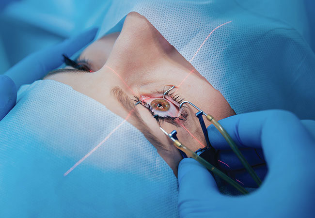
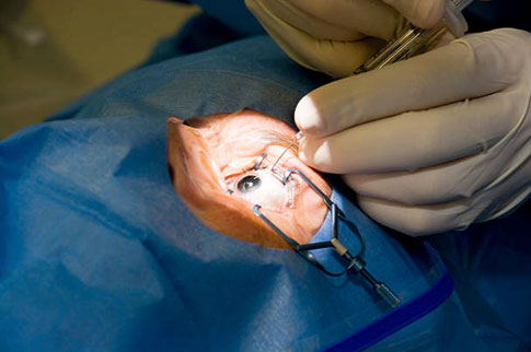
  • Numbing the eye: First local anesthetic, usually as drops or injectable agents, is provided without concern for popularity. You will be awake throughout your surgery but will experience little or no pain.
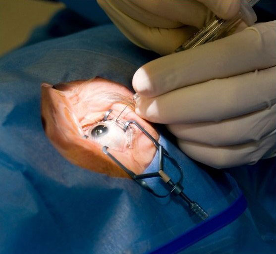
  • Tiny incision: The corneal incision (or at the junction with the sclera) is created by the surgeon, which gives access to the internal lens.
    This causes minimal trauma to the tissue, as the incision will be small and precise.
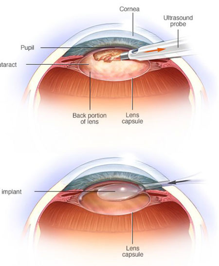
  • Breaking the cloudy lens (Phacoemulsification or other methods) : In many cases, ultrasonic energy or gentle mechanical techniques are used to fragment the cloudy natural lens. These fragments are then removed carefully via suction.
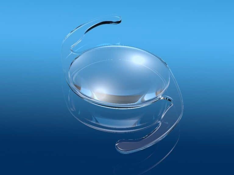
  • Implanting an artificial lens (IOL): A clear intraocular lens is inserted into the same position where the natural lens used to lie.
  • Wound closure and protection: The commonly self-sealing site injuries need no stitches. A protective shield or patch is put over the eye temporarily.

It is because of these precise steps and instrumentation of modernity that the surgery is exceedingly well tolerated.

What Symptoms You May Experience During the Surgery

There is no pain; rather, these sensations would be considered normal:-

  • Pressure or mild pushing
  • Detection of liquid movement
  • Bright light or flashes
  • Minor tugging or stretching- without sharp pains

As your eye is numbed and you are sometimes given a mild sedative, these sensations are, as a rule, well-tolerated: nothing as sharp or throb-pounding should be sensed.

Recovery & Discomfort Following Surgery

As soon as the surgery is completed, the period following that may have vague sensations like:-

  • Gritty or sandy feeling
  • Watery or teary eyes
  • Mild irritation or pressure
  • Bright light sensitivity or glare
  • Blurry vision initially

Such symptoms have almost always been, therefore, very short-lived and resolve within a few hours to days. Painkillers used without a prescription (for instance, paracetamol or ibuprofen) usually suffice to take care of mild pain. Vision will improve with time, clearer and sharper, as the eye heals. That clarity, sharpness, and comfort improve over days and weeks after surgery.

Recovery Period: What to Expect

Here is a guide to how healing typically progresses:-

Time Period

What Happens / What to Expect

Day of Surgery

You are discharged same day. Eye may feel gritty, vision blurry. Protective glasses used.

First 1–3 Days

Initial discomfort decreases. Eye drops start. Avoid heavy work or vigorous activity.

First Week

Vision starts recovering. All routine work can be resumed with care.

2–4 Weeks

Healing takes place. Pain goes off.

4–6 Weeks

100% recovery in a large number of cases. Final vision outcomes assessed.

Full recovery can take up to several weeks depending on individual healing rates, the complexity of the surgery, and other eye health factors.

Pain Management & Minimization

Surgeons and care teams utilize various modalities to keep one comfortable: local anesthetic agents that block pain signals, sedatives or anxiolytics administered for anxiety, micro-surgical instrumentation that minimizes tissue injury, delicate surgical techniques to eliminate unnecessary stretch, postoperative care considering topical medications and shields.

Then again, with all that precautions, the bulk of patients will feel hardly anything, as real pain is an extremely rare sensation. Also, due to all of these factors, the bulk of patients will hardly have any sensation, and genuine pain will be very rare.

When to Seek Medical Attention for Pain or Complications

Most aches and pains are trivial nuisances. There are a few alarm bells, however, that should have your attention:-

  • Pain that is sharp or becoming progressively worse
  • Total loss of vision suddenly
  • Severe redness, swelling, or increased discharge
  • Flashes, floaters, or shadows?
  • The sensation of lens movement or distortion

Such symptoms may indicate complications, e.g., infection, increased eye pressure, inflammation, or lens instability. Please don’t hesitate to call your surgeon if any alarming signs are noticed.

Final Thoughts: The Reality & the Fears

Modern techniques of cataract surgery are so very minimally invasive, brief, and generally well-tolerated. Advances in anesthesia and postoperative regimen have improved the process of pain control such that for most patients, pain is no longer an issue. Most patients may experience discomfort or pressure that is easily but temporarily controlled. The key, however, remains to follow the instructions as given by the surgeon, medicate per prescription, protect the eye, and turn up for follow-up visits. With good care, you can expect improved vision with minimal discomfort on the road to recovery.

What to Do & What to Avoid After Cataract Surgery

Cataract, indeed, is one of the safest and most effective surgeries in ophthalmology. Patients have an instant improvement in their vision, and they return to a normal routine. Careful consideration of postoperative care has assured the best outcome. Below is a very comprehensive list of what to do or not do after the cataract surgery.

Dos: Best Practices for Best Recovery

  • Travel as directed – A course of eye drops is usually prescribed to all patients: antibiotics and anti-inflammatory preparations for preventing infection and lessening the swelling. Wash your hands always before any medication administration and use the custom program recommended by your surgeon. They are to be continued till the date of post-op check-ups, as prescribed.
  • Give some rest to the eye – While you may notice immediate improvement in vision, the eye needs time to heal. On those first two or three days, do not engage in very strenuous work, do any bending down, or lift heavy objects. Light distraction, like reading and watching some television, and casual use of the computer, is usually fine unless your doctor advises against it.
  • Put on your protective shield or glasses – To prevent unintentional rubbing or dust irritating the eye, go outside with protective glasses on. Also, it protects from the wind, dust, and pollen, much, etc.
  • Pain Killer when Needed – Minimal pain often felt after surgery would come with a sensation of gravel in the eye; watering and aching may occasionally follow this complaint. Most discomforts will usually be answered with a commercial pain reliever such as paracetamol or ibuprofen. Again, such medications should be used only as prescribed and in association with your prescribed eye drops.
  • Shower safely and wipe gently – One can shower or bathe after surgery, but not soap, shampoo, or let water get into the operated eye. Instead of splashing water onto oneself, allow the water to flow onto the body and gently wash one’s face with a wet cloth. For the operated eye, use cooled boiled water, cotton wool or gauze, and wipe very slowly from the inner corner to the outer corner. When wiping, never press or rub the eye.

Don’t: The Best Practices Not to Follow After Cataract Operation

  • Don’t give yourself a driving license – You are not supposed to drive yourself at all after surgery. You can drive only after the ophthalmologist has cleared you, and also when you can see legally. This may take three to four days or even longer.
  • Don’t rub or touch your eye – Even a gentle rubbing or pressure on your eye can interfere with the healing, may displace the lens, or infect it. During the first weeks of recovery, avoid touching the eye at all.
  • Not Too Soon to Reapply Eye Makeup – Particles from mascara, eyeliner, or eye shadow may float into the eye under healing and cause irritation or infection. It is generally suggested not to wear any eye makeup for at least a few weeks, with an important exception made at the end of the week until your doctor says it’s fine.
  • Don’t Expose the Eyes to Irritants – Always keep your eyes away from dust, smoke, wind, pollen, or chemical fumes. Don’t engage in activities like cooking greasy foods or doing housework that might cause splatter or limitless airborne particles.
  • Don’t Swim or Go into the Sauna – Hot tubs, saunas, and steam rooms tend to be fertile ground for bacteria and infection; hence, they should be best avoided for a few months. Usually, it’s always 4 to 6 weeks of avoidance before healing enough takes place in the eye.

Returning to Work Again and Resuming Normal Activities

  • When to resume work – Most patients return to work about three days after their operation if their jobs require little or no effort. If you are in a job requiring manual labor or extensive visual activity, you may require longer to stay off work. Follow your doctor’s advice on this matter.
  • Exercising and Moving Heavy – Mild walking is generally acceptable after surgery, but things that put a strain on the body or eyes, such as lifting heavy things, or intense workouts, are usually not advised until told otherwise by your surgeon.
  • New Glasses – Your prescription might change as your eye settles. This is generally when an optometrist will see you four to six weeks after surgery to clarify whether glasses need to be replaced. It is also generally discussed after surgery in check-ups.  

When Help is needed or Red Flags to Watch For

Look out for symptoms that might mean complications and report to your surgeon or eye clinic if you notice:-

  • Increased pain, swelling, or redness
  • Suddenly worsening of vision or blurring that will not go away
  • Yellowish or greenish discharge from the eye
  • Flashes of light, floaters, or shadows in your vision
  • A sensation that the lens is shifting and distorted vision

All of these should be assessed immediately to rule out complications like infection, retinal detachment, or implant dislocation.

Final Thought

Typically, recovery from cataract surgery is rather simple and swift; however, your diligence during the postoperative days and weeks will determine your eventual results. Thus, straying from the aforementioned “dos” and “don’ts,” along with continuous communication with your eye surgeon, will give you the best chance of a clear, comfortable vision and a successful recovery process.

SICS Cataract Surgery: An Overview

Cataract (मोतियाबिंद) is one of the very common ocular abnormalities, where the natural lens of the eye becomes cloudy and causes blurring/dimming of vision. SICS (Small Incision Cataract Surgery) or ‘छोटे चीरे से मोतियाबिंद ऑपरेशन’ is one of the surgical techniques for cataract extraction. This method has many advantages, especially in resource-limited settings or where advanced Phacoemulsification may not be necessarily ideal. Further below, we will elaborate on how SICS works, what benefits or limitations it has, the collection process, recovery, and who might be acquainted with it.

What is SICS?

Small Incision Cataract Surgery” is a manual cataract surgical procedure wherein the surgeon makes a 6-7mm incision (tunnel) in the sclerocorneal region to access and remove the cloudy lens and then implant an artificial intraocular lens (IOL). The incision is larger than that of Phacoemulsification but smaller and much better controlled than that of older extra-capsular techniques.

In SICS, the nucleus (the hard core of the lens) is usually expressed manually, often with the help of hydrostatic pressure or viscoelastic fluid, and not by ultrasonic energy. The foldable or rigid IOL is positioned after lens material removal.

Why Go in for SICS? The Key Advantages

SICS enjoys some practical and clinical benefits, especially in certain contexts:-

  • Cost-effective: Less dependency on expensive machines and fewer expensive instruments may make SICS affordable.
  • Reliability in dense cataracts:Phacoemulsification may not work well in very dense or advanced cataracts, while many circumstances have demonstrated greater success with SICS over harder nuclei.
  • Reduced dependence on electricity or high technology: Very beneficial in locations where electrical supply is poor, with some intermittent power, or where resources are scarce, SICS stands well.
  • Good visual outcomes: Those performed correctly often achieve excellent postoperative vision.
  • Possibility of suture less closure:The incision is constructed such that it self-seals, thereby reducing the need for stitches but sometimes sutures may be needed.
  • Fast & Reproducible: SICS-qualified surgeons can perform the operation rather quickly, thus enhancing throughput in busy centers.

Limitations & Trade-offs

Yet, though SICS is a strong option, some trade-offs accompany it:-

  • Larger incision size than Phaco: The tunnel in SICS is larger than that in phaco, and therefore, it may produce slightly more induced astigmatism.
  • Manual manipulation risks:Because the lens is manually expressed, there exists a unique risk of damaging adjacent structures if not done carefully.
  • Healing and visual recovery: The healing period may be modestly longer than that of small-incision Phaco procedures.
  • Skill dependency: Highly dependent on the experience of the surgeon relative to manual techniques.

Surgical Process: Step by Step

This is a typical outline of how SICS carries on:-

  • Preop evaluation – Be as complete as possible in eye examination, including corneal status, retina, intraocular pressure, axial length, and biometry.
  • Anesthesia and prep – Local usually topical anaesthesia or peri bulbar block. Sterilizing the eye and letting in dilating drops.
  • Creating Incision and Tunnel – A small but well-designed incision is made, usually as a scleral-corneal tunnel, to allow access to the lens without causing undue stress to the eye.
  • Capsulorhexis / Open Lens Capsule– The anterior capsule is held open for instrument access to lens material (Capsulorhexis).
  • Nucleus Expression / Removal – Delicately brought out through the tunnel using hydrostatic forces, viscoelastic fluid injection, or manual vectis/forceps while taking care to protect the corneal endothelium.
  • Cortical Cleanup – Now that the core of the lens, i.e., the central hard lens core, has been removed, it remains to remove the softer material outside (cortical), which should be done using a cannula.
  • Intraocular Lens Implantation – Insert and appropriately position a suitable artificial intraocular lens (IOL) in the capsular bag or elsewhere.
  • Incision Closure – Hydrate the tunnel or construct it so that it seals on its own. Additional suture less wound closure techniques are also used, depending on the surgeon’s judgment.
  • Post-operative medication – Antibiotic and anti-inflammatory drops will be administered to the patient to avoid infection and control inflammation.

Recovery & Aftercare

  • Immediate post-op – The patient may be kept in the recovery area for some time. In this period, avoid rubbing or pressing the eye.
  • Medication regimen– Eye drops (antibiotic, steroid, possibly anti-glaucoma drops) need to be given, gradually tapering them down as healing goes on.
  • Regular check-ups – Regular visits for review of eye healing, IOL placement stability, and early detection of possible complications.
  • Activity Restrictions – Do not lift heavy things, bend over, or do strenuous activities during the initial phases. Avoid water, as in swimming and splashing, until approved.
  • Visual improvement timeline – Patients will notice improvement within days, but it may take a couple of weeks for full stabilization.

Who Makes a Good Candidate for SICS?

SICS is often preferred in the following situations:-

  • Patients with dense, advanced, or mature cataracts that are harder to emulsify.
  • Settings where cost or infrastructure limits the availability of high-end Phaco machines.
  • Resource-limited or rural surgical centers.
  • Eyes that have weaker corneal parameters, wherein lesser energy is advantageous.
  • Surgeons experienced in the manual technique of cataract extraction.

Complex cases of anterior segment anatomy or weak capsular support, or those with concurrent comorbid corneal conditions, would need different approaches or advanced new surgical methods.

Their Possible Complications & Precautions

Some complications still may arise with SICS, though in general, it remains safer:-

  • Wound leakage or failure of the self-sealing incision
  • Induced astigmatism due to incision architecture
  • Damage to corneal endothelial cells
  • Capsular tears or posterior capsule rupture
  • Dislocation or Decentration of IOL
  • Infection or endophthalmitis
  • Increased intraocular pressure

Thus, care in the selection of the patient, a high degree of diligence in the surgical technique, and the operation of a stringent aseptic regimen should be followed to minimize these complications.

A Comparison between SICS and Phaco: A Brief Note

  • Incision size – Phaco uses a smaller incision (micro incisions) compared to SICS.
  • Energy use –Phaco uses ultrasonic energy to break up the lens, while SICS involves manual expression.
  • Recovery and astigmatism –Phaco tends to induce less astigmatism and faster visual recovery in ideal cases.
  • Cost & infrastructure – Phaco machines and consumables are more expensive; SICS can be more accessible.
  • Suitability with dense cataracts –SICS often has an advantage with very dense or hard cataracts.

Why SICS is popular eye treatment?

In many parts of the world, particularly in developing regions, SICS still is the backbone of cataract blindness control programs. The low cost, reproducible nature, and satisfactory visual outcome of SICS rightfully place it among the best candidates serving a wide patient population.

Surgeons who are capable of SICS know they will restore sight for many needy patients who otherwise would not have access to such high-end technology.

Conclusion

Small Incision Cataract Surgery (SICS) is a well-tested surgical procedure to treat cataracts. It is a method chosen on the basis of cost, efficacy, and adaptability. While it may not shine in every single case, it remains a mighty warrior where Phacoemulsification is not available or in eyes with dense cataracts.SICS’ success is highly reliant on the surgical aptitude, meticulous planning, and aftercare provided by the surgeon. For the rightly selected candidates, it can grant them clarity of vision, lesser dependence on spectacles, and, thus, a significantly better quality of life.

Can Cataract Surgery Be Repeated?

During a period when cataract surgery becomes perhaps the happiest operation in existence, the freedom of millions of patients who get it every year after years of clear vision is when one finds that the ophthalmologist is extremely versed in cataract surgery. However, in a few cases, some patients come back several months or years later, asking, “Will the cataract surgery be repeated?”

A cataract can never be a regrowth of fresh tissue after surgery; however, a percentage of the cases with blurry vision may sometimes be experienced, or another surgical maneuver may be applied. This could allay some vexed patients’ minds, perhaps before a decision was made to go ahead with the health of those eyes once more.

What Really Happens During Cataract Surgery?

Knowing exactly what happens at the location of the operation during surgical intervention is crucial in determining the feasibility of a repeat cataract surgery. During cataract surgery, the cloudy lens of the eye is removed, and an artificial lens called an IOL is inserted. The IOL is completely transparent and designed to last a lifetime.

Since an actual lens is being removed up to present, cataract cannot return. The proteins that caused cloudy appearance become absent, and the IOL cannot develop cataracts. That is perhaps one of the most comforting things many ophthalmologists tell their patients:” Cataract surgery is performed once for every eye.”

Some Reasons Hereafter That May Cause Vision To Haze Again

Even so, cataract surgery is eternally involved; some months, often years later, mostly for cases that are afflicted with hazy vision again, as to think that the cataract would return. They are not suffering from a cataract but from Posterior Capsule Opacification (PCO)-sometimes called a secondary cataract.

The capsule will be intact during the operation, where the primary natural lens is. That capsule will support the IOL. Cells from the original lens may have lingered in this existing capsule to thicken it or cloud the vision unilaterally over time. This same blurriness of vision with the feeling of something floating in vision occurs when rays of light pass through this cloudy capsule into the visual field; thus, in essence, it is “a cataract.”

Treatments for Posterior Capsule Opacification

Posteriorcapsule Opacification is easy to treat. Typically, a rapid, outpatient, pain-free procedure called YAG laser capsulotomy is used instead of having to do another surgery to remove it. In layperson’s terms, a laser is used to make a pinhole-sized opening through the cloudy capsule, thereby allowing the inner eye to be seen clearly again.

The procedure takes a few minutes; no stitches and no anesthesia are needed. It usually gives almost instant visual clarity. Compared to cataract surgery, it is less invasive and has an extremely low risk of complications. Therefore, wherever cataracts cannot be removed, this very common effect of surgery costs hardly a minute to fix.

Cases that May Require a Rarer Reoperation

The very few shall undergo a re-cataract procedure; however, a rare case may arise when such interventions can be properly entertained.

  • Adjusting for Wrong Lens Power – Sometimes the implanted lens may not be enough for the matching power required for corrective vision. Only seldom do such minor deviations arise despite sound preoperative measurements and calculations by the surgeon, due to the unique anatomical peculiarities of each eye. If the refractive error is large enough and no reasonable amount of correction is likely to be achieved with glasses, then the IOLcan be exchanged with another one of the most suitable power.
  • Lens Dislocation or Malposition – Trauma or surgical complications at times cause this artificial lens to move from its intended position. Such instances may be complained of by the subject with double vision, halos, or distorted images, in which case the surgeon may elect to reposition the lens or to completely remove it and replace it.
  • Injury Causing Damage– A remarkable injury to the eye after cataract surgery probably damages either the implanted lens or the capsule. Depending on the severity of damage, some of these may require corrective surgery to restore vision.
  • Wish for Modern Technology Lenses onvery rare occasions, those patients with a Monofocal lens would want to be allowed to update it later to a more expensive multifocal or toric lens for better improvement in near or distance vision. Such wishes are sometimes honored, as lens exchange for lifestyle improvement remains rare.

Is It Not So Much Repeating Cataract Surgery?

“True repeat cataract operation” should be well distinguished from other procedures performed after cataract surgery. Opening up the lens affixed to the eyes is never done on that set of eyes for the second time. Lens adjustment or exchange, or treatment of associated complications with laser, may be required at some later stage.

Thus, in that light, when asking if cataract surgery can be repeated, the response is this: The cataract of the original will not come back, but some post-surgical condition will need to be assessed for the patient.

Risks & Considerations of Additional Procedures

Of course, all surgical or laser methods involve some risks, however slight they might be. Following a cataract operation, the ophthalmologists carry out evaluations before deciding to recommend such procedures. Some of the aspects include:-

  • Healing capacity:Multiple operations take longer to heal, and these are more prone to inflammation.
  • Capsule stability:The capsule that holds the artificial lens must be intact. If weakened, secondary operations become more complicated.
  • Health of the retina: Retinal conditions from the beginning bear influences on outcomes from any other intervention.
  • State of the eyes generally:General conditions such as glaucoma or corneal diseases, or previous intervention, can have implications for the safety and efficacy of yet another one.

Reassuring Facts to Patients

  • Usually, cataracts need only to be removed once for each eye. It is rarely repeated.
  • Blurry vision years down the line are usually not a cataract recurrence, but posterior capsule Opacification.
  • PCO can easily be treated with a simple laser procedure and does not require a cataract operation.
  • Rarely, however, additional surgery might be required for instances such as lens dislocation, even though these are not related to a cataract itself, but related to the need for power correction.

Conclusion

Cataract is said to be among the safest forms of surgery and the most reliable eye operation available in today’s world. Once the opacified cataractous lens is replaced with an artificially clear lens, cataracts cannot recur. However, vision may again become cloudy due to changes in the position of the artificial lens or thickening of the capsule. However, these are problems that can be resolved through effective laser treatments, such as a YAG capsulotomy, or in rare circumstances, lens exchange.

Patients must remember that cataract surgery is not performed more than once (more or less) in a person’s lifetime and that eye specialists must be followed up regularly to ensure early detection of changes occurring after the procedure, so that the problem can be fixed immediately. In general terms, cataract surgery is not usually repeated, while eye care continues. Given that timely action is taken along with the appropriate medical guidance, clear and comfortable vision can be achieved for many years following the original operation.

Clear Lens Extraction: A Glasses-Free Way of Seeing

Wearing spectacles has become an everyday activity for those above 40 years or 50 years, be it for reading, for driving, or both. Those people keep debating whether laser surgeries like LASIK might solve the problem, but in that case, many have strong refractive errors or age-related problems in focusing, and can’t fully benefit from it. Enter Clear Lens Extraction (CLE), with a few people calling it Refractive Lens Exchange (RLE), the exciting new opportunity that portends achieving spectacle-free vision. It covers what CLE is, for whom it is best suited, potential advantages, the surgical procedure, expected recovery, and possible dangers that help in understanding this life-changing option.

What Is Clear Lens Extraction?

Clear Lens Extraction involves removing the natural crystalline lens and replacing it with an artificial intraocular lens (IOL). Unlike cataract surgery, where a cloudy lens is taken out, CLE refers to removing a perfectly clear lens that just does not meet the refractive requirements of the eye anymore. It aims at refractive errors such as myopia (nearsightedness), hyperopia (farsightedness), presbyopia (age-associated decreased near vision), or astigmatism, and it attempts to reduce or provide an approximation of providing visual independence from glasses or contacts.

The procedure is quite similar in technique and instrumentation to that of cataract surgery, which means an experienced eye surgeon could consider it a fairly reliable option. The choice of IOL or intraocular lens significantly impacts personalizing the outcome according to the lifestyle and vision aspirations of the patient.

Types of Intraocular Lenses (IOLs)

Patients now have various kinds of IOLs available to them in accordance with what they require for their vision:-

  • Monofocal IOLs: These focus vision at a single distance, typically for far vision, meaning reading glasses may still be needed for close-up tasks.
  • Multifocal or Trifocal IOLs: These advanced lenses provide clear vision at multiple distances—near, intermediate (e.g., computer work), and far—often eliminating the need for glasses entirely.
  • Toric IOLs: Designed to correct astigmatism, these are available in Monofocal or multifocal versions for customized vision correction.

To select the right IOL, surgeons perform precise measurements using tools like an A-Scan and apply specialized calculations to ensure optimal lens power.

Who Is a Good Candidate?

Clear Lens Extraction is definitely not for everybody; certain clients are the best candidates:-

  • Motivated to ditch glasses : Those eager to reduce or eliminate reliance on glasses or contact lenses.
  • Age 45-75: Mostly those who have presbyopia and those who are poor candidates for laser, like LASIK.
  • High refractive errors or thin corneas: CLE is a good option for those patients who have prescription extremes or corneas that are too thin to perform LASIK or PRK.
  • Healthy eyes: They should not have certain gross problems regarding the cornea, retina, or optic nerve.
  • Overall good health : Systemic conditions such as uncontrolled diabetes or those with autoimmune disease may eliminate the possibility of CLE benefits.

Sometimes, patients with certain types of glaucoma can also obtain some benefits from CLE treatment, like primary angle-closure glaucoma, in which a very thick natural lens increases eye pressure. A thinner IOL could provide relief in this aspect. CLE isn’t for those who have had a retinal detachment, macular degeneration, or other serious eye conditions.

Advantages of Clear Lens Extraction

There are so many things that provide viability to CLE:-

  • Spectacular fast recovery of vision: Most patients notice visual improvement within hours or days.
  • Spare the cornea: Unlike LASIK, CLE does not touch the cornea; hence, it is suitable for people with thin or irregular corneas.
  • Cure for a future cataract: CLE removes the possibility of developing cataracts later by substitution of the natural lens with an IOL.
  • Results are lasting: The IOL offers stable vision for many years as long as no new conditions develop in the eye.
  • Full correction: Premium IOLs can correct distance, near, and astigmatism, all in a single procedure.
  • Alternative for LASIK-ineligible patients: Achieving clear vision is still possible in high prescriptions or corneal limitations.

The Surgical Procedure

The CLE procedure very much resembles that of cataract surgery and is executed very precisely as follows:-

  • Anesthesia – Either topical drops or local anesthesia keeps the patient comfortable.
  • Small incision – The cornea is opened with a tiny incision (less than 2 mm).
  • Lens removal – Phacoemulsification is used to pulverize the natural lens, suctioning it out in a gentle manner.
  • IOL placement – An incision is created through which a foldable IOL is inserted and securely positioned within an eye.
  • Self-sealing incision – No stitches are needed, as the small incision closes naturally.

About 10 to 15 minutes are needed for each eye during the procedure; usually, the second eye will be operated on in a few days so that the healing process of the first one can be observed.

Recovery & Postoperative Care

Most patients return home the same day and undergo rapid recovery. Vision usually begins to improve in some cases within hours, with complete vision gradually improving over several weeks as the brain adapts to the new lens. After surgery, they go through the following measures:-

  • Medicated eye drops, including antibiotic and anti-inflammatory drops, to prevent infection and reduce swelling.
  • Activity restrictions, such as avoiding rubbing their eyes, swimming, or heavy lifting for a short period.
  • Follow-ups with regular check-ups over the course of one month to evaluate the healing process and stability of the lens.
  • Most normal activities, like reading or screen time, are resumed in one or two days.

With proper care, patients mostly enjoy sharp vision, have stable sight, and experience smooth recovery.

Risks & Complications

Although CLE is considered safe, it has risks similar to cataract surgery:-

  • Retinal detachment is more common in highly nearsighted patients.
  • Infection happens, though rarely, with serious consequences for the eye if it does occur.
  • Growth of glaucoma may cause increased eye pressure in some patients.
  • Visual disturbances: glare, halos, and reduced contrast sensitivity may occur with multifocal IOLs.
  • Posterior capsule Opacification: clouding of the lens capsule, which can easily be fixed using a laser.
  • Documented rare instances of lens misalignment where the IOL slipped and needed adjustment.

Careful screening before surgery and diligence in following post-operative precautions greatly reduce the incidence of these risks.

Summary & Outlook

Surgery by CLE is the most excellent solution for those who want liberation from glasses or contact lenses. It does so by replacing the natural lens of the eye with an individualized intraocular lens correcting, at one go, myopia, hyperopia, presbyopia, and astigmatism, making it most applicable to people unsuitable for LASIK/PRK as well as for those wishing to minimize any future cataract surgery.

Although not suitable for everyone, CLE has a quick recovery, long stability, and the chance to be free of spectacles. An in-depth consultation with an eye specialist should discuss suitability, lens options, and measures to align expectations with potential outcomes. When suitable, CLE changes lives by enabling clear, unaided vision for many years.

मोतियाबिंद (Cataract) के प्रकार, लक्षण और उपचार

क्या है मोतियाबिंद?

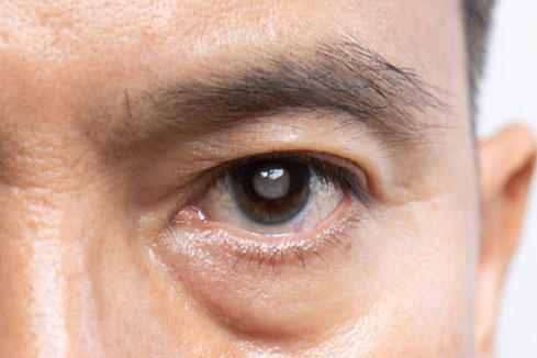
मोतियाबिंद एक नेत्र संबंधी समस्या है जिसमें नेत्र (आँख) की लेंस (crystalline lens) की पारदर्शिता धीरे-धीरे कम हो जाती है, और व्यक्ति की दृष्टि धुंधली होने लगती है । यह प्रक्रिया आमतौर पर समय के साथ होती है और एक या दोनों आँखों को प्रभावित कर सकती है। शुरुआत में व्यक्ति शायद फर्क महसूस न करे, लेकिन जैसे-जैसे रोग बढ़ता है, दृष्टि प्रभावित होती है और उसकी दैनिक गतिविधियों में बाधा आने लगती है।
मोतियाबिंद के लक्षण

आमतौर पर मोतियाबिंद के निम्नलिखित लक्षण व्यक्तियों में देखने को मिल सकते हैं:-

  • दृष्टिधुंधली या अस्पष्ट हो जाना
  • रातमें या धीमी रौशनी में देखने में कठिनाई
  • प्रकाशके इर्द-गिर्द हल्की आभा (halo) दिखना
  • तेज़धूप या सूरज की रौशनी में चुभन या परेशानी
  • पढ़तेसमय या अन्य सूक्ष्म कार्यों के लिए अधिक रोशनी की आवश्यकता
  • चश्माया कॉन्टैक्ट लेंस की क्षमता में लगातार बदलाव
  • रंगफीके या पीले लगने लगना
  • एकही आँख में दोहरी दृष्टि

ये सभी संकेत यह इशारा करते हैं कि लेंस की स्थिति बदल रही है और जल्द से जल्द नेत्र विशेषज्ञ से जाँच करानी चाहिए।

मोतियाबिंद के कारण

मोतियाबिंद विकसित होने के कई कारण हो सकते हैं, जिनमें से कुछ प्रमुख निम्नलिखित हैं:-

आयु(Age)

जैसे-जैसे उम्र बढ़ती है, लेंस के प्रोटीन संरचना में परिवर्तन आना स्वाभाविक है जो समय के साथ लेंस की पारदर्शी बनाए रखने की क्षमता को कमजोर कर देते हैं।

धूम्रपान(Smoking)

धूम्रपान करने वालों में विशेष रूप से न्यूक्लियर या पोस्टरियर सबकैप्सुलर प्रकार का मोतियाबिंद विकसित होने का खतरा बढ़ जाता है।

पराबैंगनी (UV) विकिरण

यदि आपकी आँख सीधे सूर्य की UV किरणों के संपर्क में अधिक समय तक रहती हैं, तो लेंस कोशिकाओं को नुकसान पहुँच सकता है, जिससे मोतियाबिंद की संभावना बढ़ जाती है।

दीर्घकालिक दवाओं का उपयोग

विशेष रूप से कॉर्टिकोस्टेरॉइड और कुछ अन्य दवाएँ लेंस को प्रभावित कर सकती हैं और मोतियाबिंद की समस्या उत्पन्न कर सकती हैं।

नेत्रीय चोट या आघात

अगर आँख को चोट लग जाए — आगे की लेंस कैप्सूल प्रभावित हो जाए — तो लेंस की पारदर्शिता बाधित हो सकती है।

अन्य स्वास्थ्य स्थितियाँ

मधुमेह जैसे रोग, उच्च रक्तचाप, पोषण की कमी आदि भी लेंस को प्रभावित कर सकते हैं और मोतियाबिंद की प्रक्रिया को तेज कर सकते हैं।

मोतियाबिंद के प्रकार

मोतियाबिंद कई तरह के होते हैं, जिनका वर्गीकरण लेंस की स्थिति और मोतियाबिंद के स्थान के आधार पर किया जाता है। नीचे प्रमुख प्रकार दिए गए हैं:-

सबकैप्सुलर मोतियाबिंद(Subcapsular Cataract)

इस प्रकार लेंस की पृष्ठीय (पिछली) सतह के नीचे विकसित होता है, जो लेंस को घेरे रहने वाली कैप्सूल के अंदर की ओर होता है। ऐसी स्थिति विशेष रूप से उन लोगों में अधिक देखी जाती जाता है जो स्टेरॉयड दवा लेते हैं या मधुमेह से प्रभावित हैं।

न्यूक्लियर स्क्लेरोटिक मोतियाबिंद (Nuclear Sclerotic Cataract)

यह बेहद आम समस्या है, जो ख़ास तौर पर उम्र बढ़ने के साथ जिसमे लेंस का केंद्र (न्यूक्लियस) कठोर और पीला होकर धुंधला हो जाता है।

कॉर्टिकल मोतियाबिंद(Cortical Cataract)

यह लेंस के बाहरी भाग (कॉर्टेक्स) में सफेद, धुंधले “स्पोक जैसे” निशान बनाता है जो केंद्र की ओर बढ़ते हैं और प्रकाश को केंद्र तक पहुँचने से रोक सकते हैं।

मोतियाबिंद का इलाज / सर्जरी

मोतियाबिंद का मुख्य एवं प्रभावी इलाज सर्जरी (मोतियाबिंद निकालने की प्रक्रिया) ही माना जाता है। आइए इस प्रक्रिया और उसके बाद की देखभाल पर एक नज़र डालें :-

सर्जरी से पहले की तैयारी

  • आपकानेत्र चिकित्सक आपके आँखों की बारीकी से जाँच करेगा और यह तय करेगा कि किस प्रकार का कृत्रिम लेंस (IOL) आपके लिए सबसे ज्यादा उपयुक्त होगा।
  • आपकोनिर्देश दिए जाएंगे कि सर्जरी से पहले क्या तैयारी करना है—जिसमे कुछ दवाओं का प्रयोग छोड़ना, आँखों की स्वच्छता बनाए रखना आदि शामिल हैं।
  • सर्जरीकी विधि
  • यहएक दिनचिकित्सा (outpatient) प्रक्रिया हो सकती है, जो लगभग एक घंटे या उससे कम समय में पूरी हो जाती है।
  • शुरुआतमें आँख की पुतली को फैलाने वाली दवाएं दी जाती हैं और एनेस्थीसिया (स्थानीय बेहोशी) की प्रक्रिया पूरी की जाती है।
  • धुंधलेलेंस को सावधानीपूर्वक निकालकर उसकी जगह एक कृत्रिम लेंस (IOL) स्थापित कर दिया जाता है।
  • सर्जरीके बाद बहुत छोटे आकार का चीरा (incision) रह सकता है, जिसे आमतौर पर टैग (stitch) की आवश्यकता न हो।

सर्जरी के बाद की देखभाल

  • कुछसमय तक एंटीबायोटिक व स्टेरॉयड आई ड्रॉप्स का उपयोग करना आवश्यक होता है।
  • आँखको रगड़ने या दबाने से बचें।
  • सामान्यगतिविधियाँ धीरे-धीरे शुरू करें, लेकिन भारी व्यायाम, पानी में जाना या आँखों में झटका देने वाले काम पहले कुछ सप्ताह से बचें।
  • नियमितरूप से डॉक्टर के पास जाँच कराते रहें ताकि यह देखा जा सके की रोगी की स्थिति ठीक हो रही हो या नहीं ।

संभावित जटिलताएँ

  • संक्रमणया सूजन
  • आँखमें दबाव बढ़ जाना (ग्लूकोमा जैसा लक्षण)
  • कृत्रिमलेंस का अपनी जगह से खिसक जाना (डिसलोकेशन)
  • पीछेकी लेंस कैप्सूल का बाद में धुंधलापन (posterior capsule opacification), जिसे लेज़र द्वारा ठीक किया जा सकता है
  • कभी-कभीरेटिना अलगाव जैसी गंभीर समस्या

मोतियाबिंद से बचाव के उपाय

मोतियाबिंद को पूरी तरह से रोक पाना आसान नहीं है, लेकिन उसकी शुरुआत को धीमा जरूर किया जा सकता है जिसके लिए निम्नलिखित सावधानियाँ बेहद उपयोगी हैं:-

नियमित आँखों की जाँच कराएँ

विशेष रूप से 40 वर्ष से बाद नियमित आँखों की जांच और विश्लेषण आवश्यक है। इससे मोतियाबिंद या दूसरे नेत्र रोगों को शुरुआती दौर में पहचानना आसान हो जाता है।

संतुलित और पोषण युक्त आहार लें

विटामिन C, E, ज़ीनथिन, ल्युटिन तथा ओमेगा-3 जैसे तत्वों से भरपूर भोजन लें — जैसे हरी पत्तेदार सब्जियाँ, मछली, अंडे आदि।

धूम्रपान बंद करें

धूम्रपान लेंस में ऑक्सीडेटिव तनाव बढ़ाता है, जिससे मोतियाबिंद विकसित होने की संभावना बढ़ जाती है।

शराब का सेवन बंद या सीमित करें

अत्यधिक शराब स्वास्थ्य को प्रभावित कर सकती है और मोतियाबिंद के जोखिम को बढ़ा सकती है।

सूर्य क ेतेज प्रकाश से बचें

बाहर निकलते समय UV सुरक्षा चश्मे और टोपी का उपयोग करें ताकि आँखों को हानिकारक पराबैंगनी किरणों से बचाया जा सके।

मधुमेह और अन्य बीमारियों का नियंत्रण रखें

यदि आप मधुमेह, उच्च रक्तचाप जैसी स्थितियों से ग्रस्त हैं, तो उन्हें नियंत्रित रखना आवश्यक है, ताकि वे आँखों को हानी न पहुँचाएँ।

निष्कर्ष

मोतियाबिंद एक सामान्य लेकिन दृष्टि को प्रभावित करने वाली समस्या है, जो समय के साथ बढ़ती है। शुरुआत में धुंधलापन हल्का हो सकता है, लेकिन धीरे-धीरे यह जीवन की गुणवत्ता को प्रभावित कर सकता है। आधुनिक नेत्र सर्जरी (IOL प्रत्यारोपण सहित) एक सुरक्षित, प्रभावशाली और स्थापित इलाज है। सही समय पर नेत्र विशेषज्ञ से परामर्श लेकर आप बेहतर उपाय कर सकते हैं और अपनी दृष्टि को बनाए रख सकते हैं।

कैसे करें मोतियाबिंद से बचाव?

आँखें हमारी जीवन की अमूल्य अमानत/धरोहर हैं जो हमें अच्छी दृष्टि दे कर हमारे तमाम रोजमर्रा के कामों, जैसे कि पढ़ने-पढ़ाने, ऑफिस के कार्य, एवम अन्य गतिविधियों को पूरा करने में अपररिहर्य योगदान करती हैं । लेकिन बढ़ती उम्र और आधुनिक जीवनशैली की चुनौतियाँ (जैसे स्क्रीन समय बढ़ जाना, प्रदूषण, अस्वस्थ भोजन आदि) के चलते हमें अनेक विकृतियों का सामना करना पड़ता है । इनमें से एक सबसे सामान्य लेकिन गंभीर समस्या है मोतियाबिंद (Cataract), जिसको एक समय के भीतर उचित कदम उठाते हुए रोका जा सकता है । हम यहां पर कुछ ख़ास उपाय और जानकारी आपके साथ साझा करने जा रहे हैं जो आँखों को स्वस्थ रखने रखने के साथ साथ मोतियाबिंद की समस्या से बचने में भी आपकी मदद करेंगे:-
मोतियाबिंद क्या है और क्यों होता है?

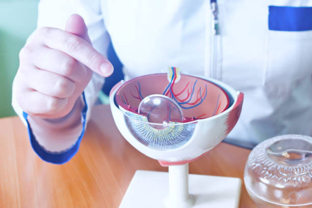
आइए सबसे पहले जानते हैं कि मोतियाबिंद होता क्या है। दरअसल ये समस्या तब उत्पन्न होता है जब आंख की लेंस (lens) पारदर्शिता खोने लगती है और धीरे-धीरे ये धुंधली या बिलकुल गायब सी हो जाती है। यह स्थिति आँखों के लेंस में मौजूद प्रोटीनों का आपस में चिपक जाने या वहां के अन्य जैविक रासायनिक प्रक्रियाओं की वजह से उत्पन्न होती है जो उम्र बढ़ने के साथ यह स्वाभाविक रूप से और भी तेज़ हो सकती है। लेकिन उम्र ही एक मात्र कारण नहीं है। इसके बढ़ने के दूसरे कारण हो सकते हैं जो निम्नवत:-

  • अत्यधिकपराबैंगनी (UV) किरणों का संपर्क
  • धूम्रपान, शराबका सेवन
  • मधुमेह, उच्चरक्तचाप जैसी बीमारियाँ
  • दीर्घकालीनस्टेरॉयड दवाओं का उपयोग
  • आनुवांशिक(hereditary)

इन कारकों की समझ और नियंत्रण ही हमें इस समस्या से बचने की दिशा में प्रारंभिक सहायता प्रदान करते हैं।

मोतियाबिंद से बचने के उपाय — अपनाएँ ऐसी आदतें

नीचे कुछ व्यावहारिक और सहज उपाय दिए गए हैं, जो नियमित रूप से अपनाने पर आपकी आँखों को सुरक्षित रख सकते हैं:-

धूप और पराबैंगनी किरणों (UV) से बचाव

सूर्य की किरणों में उपस्थित UV-A और UV-B किरणें आँखों की कोशिकाओं को ऑक्सीडेटिव तनाव पहुंचा सकती हैं। यह लेंस में फ्री रैडिकल की उपस्थिति बढ़ा सकती है और प्रोटीनों को नुकसान पहुंचा सकती है।

इसलिए जब भी बाहर जाएँ — खासकर दोपर में तो यूवी फिल्टर वाले धूप के चश्मे और टोपी का प्रयोग जरूर हानिकारक करें ताकि आँखों तक हानिकारक UV पहुँच न सके।

संतुलित पौष्टिक आहार

आँखों की सेहत के लिए भोजन की भूमिका भी अनिवार्य है जिसके अंतर्गत निम्नलिखित पोषक तत्व विशेष रूप से लाभदायक हैं:-

  • एंटीऑक्सीडेंट्स(जैसे विटामिन C, विटामिन E)
  • कारोटेनॉइड्स(जैसे ल्यूटिन, ज़ीएक्सैंथिन)
  • हरीसब्जियाँ (पालक, मेथी आदि)
  • रंगीनफल (जामुन, संतरा, आम आदि)
  • ओमेगा-3 फैटीएसिड (मछली, अखरोट आदि में)
  • प्रोसेस्डया तली हुई चीजों के बजाय ताज़ी और प्राकृतिक भोजन को तरजीह दें।

आँखों का चोट से बचाव

खेलकूद, घरेलू काम या अन्य गतिविधियों से आँखों पर अनजाने में चोट लग सकती है जो बाद में आपकी आँखों के लेंस को नुक़सान पहुंचा सकती हैं।

  • खेलतेसमय चश्मे पहनें
  • रसायनोंया धूल वाले माहौल में सुरक्षा गॉगल्स का उपयोग करें

नियमित आंखों की जाँच

समय-समय पर किसी नेत्र विशेषज्ञ//चिकित्सक (ऑप्टोमेट्रिस्ट) से जाँच करवाते रहें, खासकर जब आप 40 वर्ष को पार चुके हों या अपनी आँखों में आप धुंधलापन महसूस करने लगे हों।

ऐसी जाँच में शुरुआत में ही मोतियाबिंद या दूसरी आँखों की समस्या पकड़ में आ सकती है, जिससे उसे नियंत्रित करना और उसका निदान बेहद आसान हो जाता है।

जोखिम और सावधानियाँ

अधिक UV एक्सपोजर – धूप या उच्च ऊँचाई वाले स्थानों पर UV स्तर अधिक होता है। यदि नियमित सुरक्षा न की जाए, तो लेंस की कोशिकाएँ आनुवांशिक क्षति का शिकार हो सकती हैं।

धूम्रपान व शराब – ये आदतें शरीर में ऑक्सीडेटिव तनाव को बढ़ाती हैं और आँखों की रक्षा प्रणाली को बेहद कमजोर कर सकती हैं।

मधुमेह / उच्च रक्तचाप – बेहद नियंत्रणहीन रक्त शर्करा या रक्तचाप आँखों की सूक्ष्म रक्तवाहिकाओं को क्षति पहुंचा सकते हैं, जो लेंस पर भी साकार हो सकती है।

दवाईयों का दीर्घकालीन उपयोग – खासकर स्टेरॉयड दवाओं का लंबे समय तक उपयोग मोतियाबिंद की प्रक्रिया को बढ़ा सकता है। यदि आप इन दवाओं की नियमित सेवन करते हैं तो अपने नेत्र चिकित्सक से अपनी आँखों की सुरक्षा को लेकर चर्चा अवश्य करें।

पारिवारिक इतिहास – यदि परिवार में किसी को पहले ही मोतियाबिंद की समस्या रही है, तो ये भी एक जोखिम का कारन बन सकता है। इस स्थिति में और भी ज्यादा सतर्कता कि आवश्यकता होती है।

निष्कर्ष

मोतियाबिंद उम्र बढ़ने के साथ होने वाली एक सामान्य समस्या जरूर है, लेकिन जरूरी नहीं कि आपको भी इस समस्या से गुजरना ही पड़े। वक़्त रहते उठाए गए सुरक्षा कदम, स्वस्थ जीवनशैली, नियमित जाँच और आँखों की सुरक्षा के प्रति जागरूकता इस स्थिति को काफी हद तक टाल सकते हैं और इसके अन्य लक्षणों को आने वाले दिनों में एक लम्बे समय तक रोक सकते है।

यदि आप या आपके परिजन दृष्टि संबंधी नेत्र सम्बन्धी कोई समस्या महसूस कर रहे हों या मोतियाबिंद का ऑपरेशन करा चुके हैं, तो समय रहते नेत्र विशेषज्ञ से सलाह लेना न भूलें। याद रखिये शुरुआत में ही समस्या का निदान प्रभावी साबित होता है।

मोतियाबिंद सर्जरी: सावधानी ही एक मात्र उपाय

मोतियाबिंद यानी Cataract सर्जरी आज के युग की एक आम और सुरक्षित नेत्र-चिकित्सा की विशेष प्रक्रिया मानी जाती है। जिसके अंतर्गत धुंधला लेंस हटाकर उसमें स्पष्ट इन्ट्राओक्यूलर लेंस (IOL) लगा दिया जाता है जिसके बाद ज्यादातर लोगों की दृष्टि कुछ ही समय के भीतर बेहतर हो जाती है। लेकिन सफल सर्जरी के बाद भी रिकवरी का समय बेहद संवेदनशील होता है जिस दौरान अगर आवश्यक सावधानियों का निर्वहन नहीं किया गया, तो आपको कुछ जटिल समस्याओं का सामना भी कर पड़ सकता है ।

इस लेख में हम विस्तार से जानेंगे कि सर्जरी के बाद किन बातों का हमें विशेष ध्यान रखना चाहिए, किन गतिविधियों से बचना चाहिए, और अपनी आँखों की रक्षा हम कैसे करें ।

सर्जरी के तुरंत बाद: यानि की पहले 7 दिन (Initial Recovery Phase)

सर्जरी के शुरू के कुछ दिन अत्यंत महत्वपूर्ण होते हैं। इस दौरान आँखों में सूजन, हल्की लालिमा, पानी आना, या थोड़ी धुंधली दृष्टि जैसी तकलीफें सामान्य है । फिर भी इन्हें नियंत्रित करने और इनके संक्रमण से बचने के लिए ये उपाय बेहद ज़रूरी हैं:

  • डॉक्टर द्वाराबताई गयी दवाएँ समय पर लें — एंटीबायोटिक या एंटी-इंफ्लेमेटरी आई ड्रॉप्स
  • आँखोंको रगड़ने या दबाने से परहेज़ करें — खासकर सोते समय
  • नहातेसमय आँखों पर पानी न जाने दें — कम से कम एक हफ़्ते तक
  • धूप , धूल और प्रदूषण से बचाव करें — बाहर निकलते समय धूप के चश्मे पहनें
  • भारी काम न करें — झुकने, भारी सामान उठाने, और ज़ोरदार शारीरिक व्यायाम आदि से बचें
  • मेकअप, क्रीमया कॉस्मेटिक आदि बिलकुल न लगाएं — कम से कम 2–3 हफ़्ते तक

इन उपायों का पालन करने से शुरुआती दिन बेहद सुरक्षित और सहज हो जाते हैं, और आगे की रिकवरी और भी आसान हो जाती है।

  • 1–4 हफ़्ते सावधानी , जब दृष्टि सुधरने लगे : जब शुरुआती जलन और सूजन सामान्य होने लगे और डॉक्टर्स दवाइयाँ धीरे-धीरे बंद करने की सलाह देने लगें, तब ये स्टेज आ जाती है। इस दौरान भी निगरानी और संयम की सख्त जरूरत होती है :
  • दैनिक गतिविधियों को धीरे -धीरे सामान्य करें : हलाकि हल्के काम जैसे टहलना, और पढ़ना आदि शुरू कर सकते हैं
  • आँखों पर अधिक तनाव न डालें : लंबे समय तक मोबाइल, टीवी स्क्रीन आदि बचें
  • स्वस्थ आहार लें : जिसमे विटामिन, ओमेगा-3, हरी सब्जियाँ व फल आदि प्रचुर मात्रा में हों
  • नियमित चेकअप : डॉक्टर द्वारा निर्धारित फॉलो-अप पर जाएँ

इस बीच यदि आंखों में तेज दर्द होना, दृष्टि का तेजी से कमजोर होने लगना, लालिमा बढ़ना या किसी असामान्य अनुभव के होने पर तुरंत विशेषज्ञ से संपर्क करें।

चेतावनियाँ जिन्हें नज़रअंदाज़ बिलकुल न करें

कई बार सावधानी के बावजूद भी कुछ समस्याओं का सामना कर पड़ सकता है जिनमे ये लक्षण शामिल हैं जिन्हे हल्के में बिलकुल न लें:-

  • आँख मेंतेज़ दर्द या लगातार जलन
  • अचानकदृष्टि का कमजोर पड़ जाना या दोहरी दृष्टि (Double View )
  • लालिमाया सूजन का बढ़ जाना
  • प्रकाशचमक या फ्लोटर्स का बनना

इन लक्षणों को तुरंत डॉक्टर को दिखाना चाहिए — क्योंकि ये संक्रमण, रेटिना डिटेचमेंट या अन्य किसी अन्य गंभीर समस्या के संकेत हो सकते हैं।

दीर्घकालिक देखभाल : देखने की क्षमता को सुरक्षित रखना

जब प्राथमिक रिकवरी का चरण सफलतापूर्वक पूरा हो जाए, तब भी आँखों की देखभाल पूरी तरह खत्म नहीं होती। नीचे कुछ दीर्घकालिक सुझाव हैं:

  • नियमित नेत्र-परिक्षण: हर6–12 महीने में आँखों की जांच
  • चश्माया दृष्टिसुधार: यदि आवश्यक हो तो नया प्रिस्क्रिप्शन चश्मा लगाए
  • सूर्यकी तेज रोशनी से सुरक्षा: UV रोधी चश्मे, धूप से सावधानी
  • स्वस्थजीवनशैली अपनाएं: धूम्रपान बंद करें, संतुलित आहार लें, और हाइड्रेशन के लिए ज्यादा से ज्यादा से पानी पियें

इन आदतों को अपनाकर आप अपनी दृष्टि को आने वाले लम्बे समय तक सुरक्षित रख सकते हैं।

निष्कर्ष : सावधानी से दृष्टि की रक्षा

मोतियाबिंद सर्जरी यद्यपि एक प्रभावशाली उपाय है, लेकिन उसके बाद की देखभाल आँखों की दृष्टि को और भी सुदृढ़ बनाती है। शुरुआत में अगर ध्यान नहीं दिया गया तो छोटी-छोटी जटिलताएँ भी बड़ी समस्या बन सकती हैं।

इसलिए इन निर्देशों का पालन अवश्य करें:-

सफल सर्जरी ≠ यकीनन सफल जीवन ; देखभाल ही जीवन भर सुरक्षा है।

यदि आप मोतियाबिंद सर्जरी कराने वाले हैं या हाल ही में करवा चुके हैं , तो ये सारे सुझाव आपके लिए अमूल्य हो सकते हैं। आपकी जल्द स्वस्थ दृष्टि की कामना करता हूँ।

Ojas Eye Hospital A Center of Excellence for Contoura Vision, Femto Bladefree Lasik in Mumbai, India.